File:Dino.jpg
From MicrobeWiki, the student-edited microbiology resource
Dino.jpg (366 × 490 pixels, file size: 27 KB, MIME type: image/jpeg)
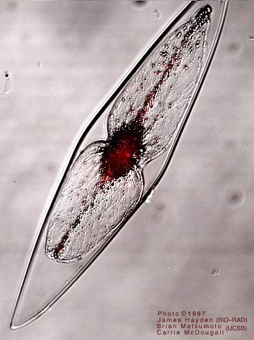
Figure 1. Pyrocystis fusiformis. The red glow is chlorophyll fluorescence (visualized with two-photon excitation microscopy) which has been superimposed over a view of the whole cell. Source: http://www.lifesci.ucsb.edu/~biolum/organism/pictures/dino.html
File history
Click on a date/time to view the file as it appeared at that time.
| Date/Time | Thumbnail | Dimensions | User | Comment | |
|---|---|---|---|---|---|
| current | 20:36, 19 April 2009 |  | 366 × 490 (27 KB) | Foflonke (talk | contribs) | Figure 1. Pyrocystis fusiformis. The red glow is chlorophyll fluorescence (visualized with two-photon excitation microscopy) which has been superimposed over a view of the whole cell. Source: http://www.lifesci.ucsb.edu/~biolum/organism/pictures/dino.html |
You cannot overwrite this file.
File usage
The following page uses this file:

