Countermeasures of Bacillus anthracis use as a Biological Weapon
Biological Weapon Overview

Biological Weapons with varying degrees of lethality have been used for many centuries and deliver toxic classifications of microorganisms such as bacteria and viruses in such a manner that causes widespread disease among many different living organisms. While intentional infection of animals and agriculture can be fear-inducing and economically detrimental, it is the infliction of disease within the human race itself that can have an extremely dramatic effect on a country’s ability to function. The use of such weapons started off in a rather elementary and blunt fashion. For example, in 1346, citizens of Kaffa (Located on the Crimean Peninsula) were attacked by the invading Tartar army via exposure to plagued corpses leading to widespread of illness.[2] We have now begun to experience much more subtle, but increasingly efficient uses of biological weapons, as displayed in the anthrax letter attack on multiple media and news offices and two United States senators in 2001, where 17 people were infected and five were killed as a result of exposure to anthrax spores(Image 1). [3]
While some may underestimate the possible impact of biological weapons relative to that of other weapons of mass destruction, such attacks should not by any means be taken lightly. A large factor as to why people do not necessarily see the potential for danger is due to the fact that the impact is more often than not initially undetectable, and even the full potential of exposure can be delayed since most weapons of this nature can live outside of a host organism for long periods of time while dormant. As such, biological weapons can be transmissible and can travel from one infected person to another; perpetuating the spread of the virus. In conjunction, these pathogens require a certain incubation period that varies from one pathogenetic species to another, so it is quite likely that an infected individual can transfer the disease before they are aware that they are affected.
Clearly, it is becoming increasingly necessary to take precautious measures against the spread of these disease-spreading biological weapons. There are a couple international policies currently in place to help in the prevention of their use: The 1925 Geneva Protocol prohibits the use of biological weapons in warfare while the 1972 Biological and Toxin Weapons Convention places restrictions on production, development, and acquisition of biological agents with malicious intentions. [3] While these are legitimate means of prevention, it is equally critical if not more important to undertake means of treatment post-infection since the increasing availability of biotechnology is making the development and dissipation of dangerous microorganisms much easier than ever before.
As foreseeable, there are a seemingly endless number of microorganisms that can be used maliciously. The selection of a biological weapon is dependent on the type of attack desired, the pathogenicity of the organism, and the incubation period (amount of time observed between exposure and symptomatic illness expression) [2]. Due to the nature of biological attacks and terrorism, some toxins are more desirable to the attacker than others. One pathogen that is especially effective is anthrax, whose etiological agent is Bacillus anthracis.
Bacillus anthracis

Bacillus anthracis has developed as a biological weapon and bioterror agent as a result of this specific bacterium's toxicity in its vegetative form, and its ability to produce spores that are tolerable of many extreme environments. This bacillus became the first bacterium to be directly linked to causing disease when scientist Robert Koch acquired a pure culture and successfully produced anthrax experimentally [4]. Koch proved the causative link to the disease by implementing his widely accepted postulates, for which he was awarded the Nobel Prize in Physiology or Medicine in 1905 [5]. The following parameters were applied in the determination that Bacillus anthracis was responsible for anthrax [5]:
(1). The microbe was found in all cases of the disease, but not present in healthy individulas.
(2). The microorganism was able to be isolated from the diseased host and then grown in a pure culture.
(3). When microbe from pure culture was introduced into a healthy, susceptible host/ model the same disease was observed.
(4). When the disease from the newly infected host was cultured, the same strain was observed with the same characteristics as initially observed.
In addition to proving the causative link from the bacterium to the disease, Koch as also able to demonstrate the ability of the bacillus to for endospores.
The anthrax bacillus is a relatively large organism measuring 1-1.2 micrometers in width and 3-5 micrometers in length. This specific bacillus can be distinguished from similar bacterium such as B. thuringiensis and B. cereus by its characteristic square-shaped ends, and centrally located ellipsoidal endospore which is associated with production of an intracellular parasporal crystal (a protein crystal)[7]. B. anthracis is a Gram-positive rod with spore forming capabilities. It is these spores formed by the bacterium that are what enable this species to be effective as a biological threat if incidentally brought into contact.
Endospores
Since the bacterium in classified as a Gram-positive organism, this allows for interestingly durable spore formations. The spores produced are resistant to relatively extreme environmental conditions and because of their thick cell walls with a high amount of peptidoglycan, they have the ability to potentially exist in a dormant state for years and even decades [8]. Even when only a small number of spores are present at the time of exposure, the spores will then germinate into their vegetative state where there exists a period of very rapid growth. This rapid growth stage typically results in host fatality due to the high concentration of toxins.
Human infection can occur when the host is exposed to the spores of the bacterium via one of more of three routes: cutaneous, gastrointestinal, or inhalation. Cutaneous contraction of the disease typically occurs through open skin wounds coming into contact with another infected organism/ part of organism. This is generally the least severe form of the disease. Gastrointestinal contraction typically results from consumption of undercooked meat that contains either the vegetative form or dormant spores of the disease. When air bourne spores are inhaled, a respiratory infection can result. Gastrointestinal and respiratory infections are historically the most letal and lead to the highest mortality rates [12].
Anthrax Toxin
It was not until 1954 that the actual toxigenic properties of B. anthracis were observed. Observationally, it was observed that in most cases of animal death from the disease, 10^9 bacteria/ml were present leading scientists to conclude that strictly the large number of bacteria caused capillary blockage by a theory known as "log-jam". experimentally, on the other hand, clinical testing showed that only 3x10^6 bacteria/ml were needed to inflict death.[4] This discovery indicated that some sort of diffusible exotoxin was responsible for anthrax's level of pathogenesis; this exotoxin being the durable spore formed by the bacterium.

In a study of the neurological and physiological responses of primates to the anthrax toxin, the apparent physically causes of death of an infected organism is due to a sharp decrease in the oxygen level, which then leads to secondary shock. This secondary shock increases the vascular permeability and partial to complete loss of cortical electrical activity (EEG) is observed in the toxin-challengedT primate. A couple hours before death, another increase in EEG activity is observed in conjunction with anoxic hypertension and eventually a complete collapse of the cardiovascular system. It was concluded that the extreme effect on the cardiovascular system is a results of direct impact of the toxin in the depth of the brain relating to the respiratory system by observation of intact junction of the phrenic and neuromuscular nerve [9]. It has also been shown that death often occurs unexpectedly and rapidly as a sharp increase in the rate the level/ lethality presence in the organism during the later stages of the disease [4].
The primary virulence toxin in B. anthracis is contained in a temperature-sensitive plasmid, pX01 which contains three different antigenetic factors [4]. Each of the following are proteins with a molecular weight of about 80kDa:
Factor I: is known as the the edema factor (EF) which is a adenylate cyclase that elevates intracellular cAMP levels and is calmodulin-dependent [10]. It is an inherent adenylate cyclase and is necessary for the edema production activity of the toxin [4]. Edema is swelling as a results of fluid build up in the body's tissues [11].
Factor II: is the protective antigen (PA) and is the binding domain of the toxin and has two domains; one for the EF and one for the lethal factor [4], which will be mentioned next. This factor is essential for the effectiveness of the toxin, since it serves as the binding site of the cytoplasmic targets for the other two factors. Without, the PA factor, the other factors will not be able to recognize their cellular targets or have means of transportation into the cytoplasm of the host from the extracellular space[10] .
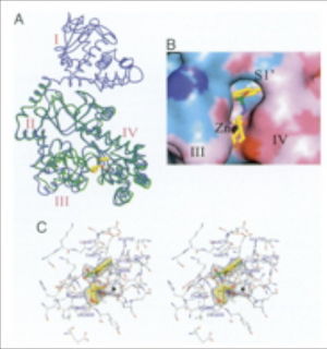
Factor III: is a zinc-dependent metalloprotease toxin known as the lethal factor (LF) [8] that targets all buMt one APK kinases [10]. Its lethality to the host cell is through the means of secretion into the host cell where it then causes signaling pathway disruptions, cell destruction, and impinges shock on the circulatory system [8]; LF is responsible for macrophage cell death.
The PA of the toxin binds to the surface of a cell recep
Section 1
Include some current research, with at least one figure showing data.
Lethal Factor Inhibition
Section 3
Include some current research, with at least one figure showing data.
Conclusion
Overall text length at least 3,000 words, with at least 3 figures.
References
[1] ["http://www.textbookofbacteriology.net/Anthrax.html"
[2] Introduction to Biological Weapons. 2010. Federation of American Scientists
[3] “Amerithrax Investigation” Federal Bureau of Investigation, 2010.
[4] [[5] Todar, K. "Bacillus anthracis and Anthrax". Todar's Online Textbook of Bacteriology. 2009.
[5] [Slonczewski, Joan L., J.W. Foster. "Microbiology: An Evolving Science". Koch's Postulates Link a Pathogen with a Disease. W. W. Norton & Company, Inc. New York, N.Y. 2009. p.21-22.]
[6] [http://srs.dl.ac.uk/Annual_Reports/AnRep01_02/anthrax-spores.jpg
[7] [Aronson, Arthur I., D.J. Tyrell, P.C. Fitz-James, and L.A. Bulla Jr. July 1982. "Relationship of the Syntheses of Spore Coat Protein and Parasporal Crystal Protein in Bacillus thuringiensis" Journal of Bacteriology Vol. 151. No.1: 399-410.]
[8] W. L. Shoop, Y. Xiong, J. Wiltsie, A. Woods, J. Guo, J. V. Pivnichny, T. Felcetto, B. F. Michael, A. Bansal, R. T. Cummings, B. R. Cunningham, A. M. Friedlander, C. M. Douglas, S. B. Patel, D. Wisniewski, G. Scapin, S. P. Salowe, D. M. Zaller, K. T. Chapman, E. M. Scolnick, D. M. Schmatz, K. Bartizal, M. MacCoss, J. D. Hermes, William C. Campbell. May 2005. “Anthrax Lethal Factor Inhibition”. Proceedings of the National Academy of Sciences of the United States of America. Vol. 102 No. 22: 7958-7963 .
[9] Klein, F., R. Lincoln, J. Dobbs, B. Mahlandt, N. Remmele, J. Walker. Feb. 1968 "Neurological and physiological Response of the Primate to Anthrax Infection". The Journal of Infection Diseases. Vol. 118 No. 1" 97-103.
[10] Abrami, Laurence, L. Shihui, P. Cosson, S. Leppla, F. Gisou van der Goot. Feb. 2003. “Anthrax toxin triggers endocytosis of its receptor via a lipid raft-mediated clathrin-dependent process”. The Journal of Cell Biology, 2003. Vol. 160: 321-328.
[11] [http://www.nlm.nih.gov/medlineplus/edema.html "Edema". United States National Library of Medicine and the National Institutes of Health. 12 April 2010.
[12] [http://www.jbc.org/content/281/8/4831.full Joyce, Joseph, C. James. Feb 2006. "Immunogenicity and Protective Efficacy of Bacillus anthracis Poly-γ-d-glutamic Acid Capsule Covalently Coupled to a Protein Carrier Using a Novel Triazine-based Conjugation Strategy" Journal of Biological Chemistry. 281: 4831-4843.
Edited by student of Joan Slonczewski for BIOL 238 Microbiology, 2010, Kenyon College.


