File:20100916 011605 Mycelium.jpg
From MicrobeWiki, the student-edited microbiology resource
Size of this preview: 600 × 600 pixels. Other resolution: 1,600 × 1,600 pixels.
Original file (1,600 × 1,600 pixels, file size: 170 KB, MIME type: image/jpeg)
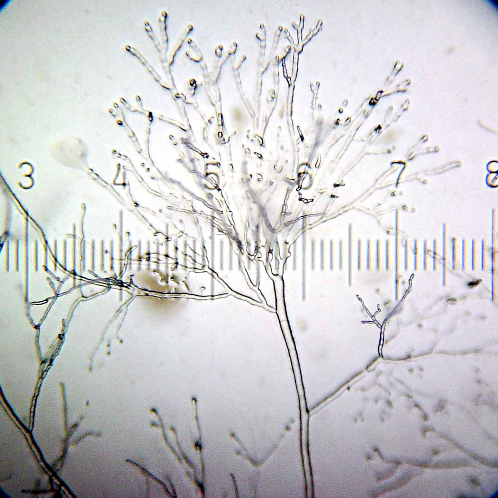
Early growth stage of an unidentified mold, grown in a Petri dish from a sample of guano from a pet dove. At this stage, only the mycelium exists. A few days later, this had grown into a very impressive fluffy white mold, that rapidly filled the Petri dish.
5× objective, 15× eyepiece; numbered ticks are 230 µM apart.
File history
Click on a date/time to view the file as it appeared at that time.
| Date/Time | Thumbnail | Dimensions | User | Comment | |
|---|---|---|---|---|---|
| current | 11:46, 18 September 2010 | | 1,600 × 1,600 (170 KB) | Blaylock (talk | contribs) | Early growth stage of an unidentified mold, grown in a Petri dish from a sample of guano from a pet dove. At this stage, only the mycelium exists. A few days later, this had grown into a very impressive fluffy white mold, that rapidly f |
You cannot overwrite this file.
File usage
The following page uses this file:
