Mimivirus
Mimivirus
[[1]]
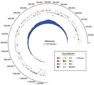
- The recently discovered giant virus Acanthamoeba polyphaga, Mimivirus, Is the largest DNA virus yet isolated (Claverie et al, 2006). Its structure, size, and genome complexity have spurred much controversy concerning the definition of a virus. Viruses have long been separated from the tree of life because of their inability to metabolize and/or reproduce independently of a host. With the discovery of more and more obligate internal parasites, both on the cellular and organismal level, and the discovery of very large and complex viruses, such as Mimivirus, the definition of a virus has been the subject of much recent debate. While a virus is traditionally defined as a non-living particle, recent discoveries about the Mimivirus genome blur the line between virus and microorganism even more, revealing astonishing complexity and an abundance of genetic material (the Mimivirus genome is 1181.4 kb long, Claverie et al, 2006).
Physical Similarities to Cellular Life
[[2]]
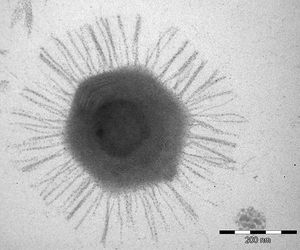
- Mimivirus is exceptional in its sheer physical size, matching small bacteria and dwarfing other viruses. The virus is so large in fact that it would not be filtered out with other, more normal sized, viruses by the filters traditionally used in lab work (Claverie et al, 2006). Viruses are often filtered out before the genetic material of the remaining bacteria/archaea/eukarya is isolated.
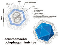
The structure of Mimivirus is so similar to that of parasitic bacterium that the particle was only identified as a virus in 2003, 11 years after its discovery in 1992 (Claverie et al, 2009). The similarities between the largest DNA viruses and the smallest microorganisms are not restricted only to size of both the particle and the genetic material. The external structure of Mimivirus is very similar to that of small prokaryotic bacteria. The icosohedral capsid of Mimivirus is coated in a layer of proteins that in some ways mimics the cell wall of Gram-positive bacteria. This protein coating is believed to act as a disguise designed to deceive the ameboid host of Mimivirus into recognizing the virion as a bacteria and compel the host to engulf the virus (Claverie et al, 2009).
The external protein coat may also serve another function. The diameter of Mimivirus without its protein coat is 0.5 micrometers. This would prove problematic for the virus, the size threshold for efficient phagocytosis is 0.6 micrometer, were it not for the protein coating, which increases the diameter of the virus particle to 0.75 micrometers. The increase in size provided by the external protein coating helps to ensure that single virion will trigger phagocytosis, and thus infection, of the host amoeba.
Genetic Similarities to Cellular Life
- Many of the same kinds of genes found in microorganisms are also found in Mimivirus and other large DNA viruses. The Mimivirus genome encodes for central components of the protein translation apparatus. The creation of these structures was thought to be characteristic of cellular organisms (Claverie et al, 2009). The isolation of these transcripts in Mimivirus further blurs the line between organism and virion. While the replication cycle of a virus would imply only the most basic and efficient suite of genes, directed towards successful virion entry, replication, and possibly avoiding the immune system, the Mimivirus genome is capable of much more than this simple replication cycle. The virus is able to induce the creation of a large and elaborate replication structure that has been described as “volcano-like” (Suzan-Monti et al, 2007). The large genome of Mimivirus also requires many chaperone genes and proteins in order to be adequately maintained.
The discovery of chaperone genes in Mimivirus has incurred hypotheses that postulate a correlation between genome size and the number of accessory genes required to maintain them. Larger genomes need more chaperone genes to ensure conservation and structural integrity of their complex genetic material. Smaller genomes do not require the same amount of maintenance, and so have fewer genes dedicated to this occupation. This hypothesis provokes another that states that as a viral genome grows, largely through the incorporation of host genetic material, it needs to evolve new methods to maintain the growing genome. Likewise, if a viral genome were to be shrinking in size, fewer genetic maintenance mechanisms would be required and those adaptations would presumably fade away, or at least become vestigial.
Phylogenetic Origin
- While many viruses are equipped with means of manipulating the internal workings of their host on a large scale, these changes are usually not as drastic as those caused by Mimivirus, and they require far fewer structures to be created and maintained. The complexity of the Mimivirus genome, and the evidence that it can be virtually indistinguishable from the genetic material of small microorganisms, forces the scientific community to rethink the definition of large DNA viruses and perhaps viruses in general. Some researchers have even hypothesized that the Mimivirus could represent an entirely new domain of the phylogenetic tree, distinct from archaea, eukarya, and bacteria. This seems like an ambitious theory to put forward, and indeed it has been shown to be not very likely (Moreira et al, 2008). A more likely explanation for the extreme complexity and size of the Mimivirus genome is that it represents a transitional organism progressing away from the traditional viral definition and towards a legitimate microorganism. Another possibility is that Mimivirus and other large DNA viruses actually arose from degenerative parasitic cells that gradually lost the ability to metabolize and reproduce independently of a host cell. Evidence for this is largely theoretical, and is based on the assumption that the transition to a parasitic lifestyle allows for a large reduction in genome size and complexity, a process that may have only affected those Mimivirus genes involved in metabolism and replication.
If it can be established that Mimivirus and other comparably large DNA viruses are a transitional phase in an evolutionary progression towards a novel lifestyle, the question becomes: towards which alternative lifestyle are these giant viruses progressing, and from where on the phylogenetic tree are they progressing? Modern evidence seems to indicate that this class of virus is descended from degenerative parasitic cells occupying a wide range of ecological niches and originating from many separate locations on the phylogenetic tree.
A sample of the twelve viruses with the largest genomes yet discovered (Claverie et al, 2006) shows that these microbes represent five distinct families of virus, many completely unrelated. This diversity, not only in the virus family of origin, but also in the host-type, vector, and ecological niche, implies that the evolution of the gigantism now observed in these viral genomes is not directed towards a specific kind of viral lifestyle. If an unusually large and complex genome were beneficial for viruses infecting a certain kind of host, or playing a certain ecological role, one would expect to find viruses with larger genomes infecting specific types of organisms and functioning in a relatively similar way.
The wide range of the viruses with large genomes suggests that they did not arise from one specific evolutionary event, but rather came to be through multiple and unrelated instances of cellular degeneration. Why a cell would evolve in such a manner as to become an obligate internal parasite is unclear. The obvious disadvantages associated with the inability either to metabolize or replicate independently offer a strong counter-argument for the adaptation to a parasitic lifestyle. There are also many benefits that a parasitic lifestyle brings. Once a parasite has infiltrated a host, it no longer needs to cope with the often harsh extracellular environment of the microbial world. The use of a larger and mature host cell as a center for replication has the advantage of making available all of the assets of the host anatomy and not restricting the potential for replication to energy accumulated by the microbe prior to infection.
If Mimivirus and other large DNA viruses did indeed evolve from living cells that degraded into the modern form, which lineage of cell did they arise from, the archaea, bacteria, or eukarya? The eukaryotes seem unlikely progenitors of Mimivirus and its relatives; the anatomical disparities are simply too great. In order for a eukaryotic cell to make the transition to a virus, it would have to lose organelles, membranes, and many other extremely complex features that would not likely be changed significantly by the point mutations common in evolution. The physical differences between large DNA viruses and other microbes, bacteria and archaea, are less significant. None of these groups have the elaborate internal membranes characteristic of eukaryotes. All three have a rigid exterior with genetic material loosely contained within, but the similarities pretty much end there. Both bacteria and archaea have a very important structure that makes them very distinct from viruses. True microorganisms contain ribosomes, the sites of translation; viruses do not, hence their obligate internal parasitic lifestyle. Ribosomes are found in all living organisms and have become a widely recognized characteristic of cellular life.
The absence of ribosome’s in Mimivirus and other large DNA viruses indicates either a long and significant transition of these degraded cells into giant viruses, a process that discarded many of the structures and abilities associated with cellular life, or another origin for this group of virus. A striking characteristic of Mimivirus is the structural similarity between its highly conserved AAAATTGA promotion motif and the TATA box core promoter element found in eukaryotes (Suhre et al, 2005). Whether the viral motif is a recently derived adaptation designed to optimize viral protein translation or an archaic form of the more recently conceived TATA box core promoter element. If this second hypothesis were true, Mimivirus would be implicated as an early progenitor of the eukaryotic domain.
Alternative Evolutionary Possibilities
- It is possible that much of the genetic material of Mimivirus and other large DNA viruses has been scavenged from their hosts over millions of years. The replication cycle of Mimivirus and many other viruses involves a great deal of interaction with the host’s genetic material. Viruses often insert their genetic material into the host’s genome. This behavior can result in the wrongful inclusion of host genetic material into a newly formed virion, or alternatively, the replication and conservation of viral genetic material on the part of the host cell. This possibility would suggest nothing more than an enduring relationship between Mimivirus and its amoeboid host.
It has been suggested that Mimivirus and other large DNA viruses may represent a domain of life separate from those traditionally discussed. Further, it has been put forward that an entirely new level of classification be utilized, one that separates living organisms with ribosomes from those without them. Both of these conjectures are extremely ambitious, and may in fact be attributable more to the desire of scientific authors to make impactful statements than to any actual experimental evidence. Mimivirus and other large DNA viruses like it have enjoyed much recent scientific attention due to their abnormal size and complexity. Physical and genetic characteristics of Mimivirus indicate that it may be an important evolutionary middle ground between viruses and cellular microbes. Much more work must be done to further our understanding of the mechanical particulars of the Mimivirus lifecycle before conclusions can be drawn about its phylogenetic origin.
Replication Cycle
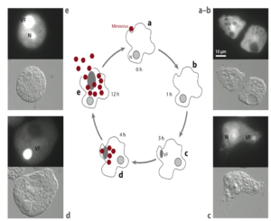
- Visual observation of Mimivirus infection shows that after 1-3 hours a distinct virus replication factory is observable. This structure continues to grow in size until about 12 hours after infection at which point new Mimivirus virions are beginning to be manufactured. Because the replication factories have been shown to match exactly the size of Mimivirus virion particles, it has been hypothesized that Mimivirus possesses transcriptional factors within its capsid. This is an adaptation observed in poxviruses, and allows the virus to begin protein synthesis immediately following entry. Such an adaptation would obviously be beneficial to Mimivirus which has so much genetic material to transcribe and duplicate.
Sputnik
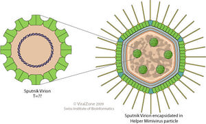
The recent discovery of virophages associated with Mimivirus further stimulates the debate over its classification as a cellular microorganism or a virus. The fact that other smaller viruses can infect Mimivirus is a true testament to its size relative to other viruses. The dependency of the virophage lifecycle on a successful infection of an amoeboid host by Mimivirus strengthens the notion of a long-standing parasitic relationship between Mimivirus and its host. The replication cycle of Mimivirus within its amoeboid host is assumed to be a long-lasting and conserved interaction, otherwise it would be very unlikely for a virophage to adapt to the specific conditions caused by Mimivirus infection.
Sputnik may act as a direct parasite of Mimivirus, exploiting the Mimivirus replication structure. It has also been proposed that Sputnik acts as a satellite parasite to Mimivirus. This implies that the smaller virus may be using Mimivirus to enter the host cell, but that its replication cycle is not dependent on Mimivirus replication structures. If Sputnik is acting as a satellite parasite its relationship to Mimivirus may not be so intimate. If Sputnik does not exploit the specific structures formed by Mimivirus infection, the smaller virus may be simply taking advantage of a convenient way to enter its ameboid host. In this case Sputnik would be considered more a parasite of the amoeba than the giant virus.
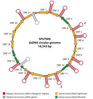
Recent evidence supports the hypothesis that Sputnik is exploiting the Mimivirus replication factories themselves, thus acting as a true virophage. In addition to the colocalization of Sputnik assembly particles with Mimivirus replication factories,[4] a hairpin structure in the genome characteristic of Mimivirus has been found in the Sputnik genome. These findings both imply that Sputnik is taking advantage of the Mimivirus replication structure and acting as a virophage.[4] There are aspects of the Sputnik genome that do not seem to support the virophage hypothesis. The Sputnik genome contains only one AAAATTGA nucleotide sequence that has been shown to act as a promoter for the transcription of Mimivirus late-early genes, indicating that the smaller virus may not be using Mimivirus structures as extensively as previously thought. Whether Sputnik is acting as a true virophage of Mimivirus or as merely a satellite virus, this relationship is very exciting and needs to be studied further.
References
Three RNA cells for ribosomal lineages and three DNA viruses to replicate their genomes: A hypothesis for the origin of cellular domain [3]
Viruses take center stage in cellular evolution [4]
Giant viruses, giant chimeras: The multiple evolutionary histories of Mimivirus genes [5]
Mimivirus and its Virophage [6]
