CTXφ Bacteriophage: Difference between revisions
No edit summary |
No edit summary |
||
| Line 12: | Line 12: | ||
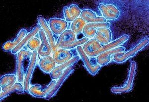
[[Image:marburgvirus.jpg|thumb|300px|right|Colony of Marburg virus. Transmission electron microscope image taken by Dr. Tom Geisbert]] | [[Image:marburgvirus.jpg|thumb|300px|right|Colony of Marburg virus. Transmission electron microscope image taken by Dr. Tom Geisbert]] | ||
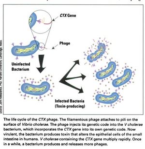
[[Image:CTX_Phage_life_cycle.jpg|thumb|left|<b>Figure 1:</b> The life cycle of the CTXφ Bacteriophage with <i>Vibrio cholerae</i> as its host. <br> Link: wordpress.com/2020/04/25/a-bacteriophage-makes-v-cholera-a-killerbug/]] | [[Image:CTX_Phage_life_cycle.jpg|thumb|left|<b>Figure 1:</b> The life cycle of the CTXφ Bacteriophage with <i>Vibrio cholerae</i> as its host. <br> Link: https://wordpress.com/2020/04/25/a-bacteriophage-makes-v-cholera-a-killerbug/]] | ||
<br>At right is a sample image insertion. It works for any image uploaded anywhere to MicrobeWiki. The insertion code consists of: | <br>At right is a sample image insertion. It works for any image uploaded anywhere to MicrobeWiki. The insertion code consists of: | ||
Revision as of 02:05, 10 December 2020
Overview
The CTXφ bacteriophage is a lysogenic, filamented phage responsible for turning the previously non-infectious Vibrio cholerae into a highly pathogenic microbe that causes disease in humans.
Select a topic about genetics or evolution in a specific organism or ecosystem.
The topic must include one section about microbes (bacteria, viruses, fungi, or protists). This is easy because all organisms and ecosystems have microbes.
Compose a title for your page.
Type your exact title in the Search window, then press Go. The MicrobeWiki will invite you to create a new page with this title.
Open the BIOL 116 Class 2019 template page in "edit."
Copy ALL the text from the edit window.
Then go to YOUR OWN page; edit tab. PASTE into your own page, and edit.
Infection, Replication & Lysing of Host Cell

Link: https://wordpress.com/2020/04/25/a-bacteriophage-makes-v-cholera-a-killerbug/
At right is a sample image insertion. It works for any image uploaded anywhere to MicrobeWiki. The insertion code consists of:
Double brackets: [[
Filename: PHIL_1181_lores.jpg
Thumbnail status: |thumb|
Pixel size: |300px|
Placement on page: |right|
Legend/credit: Electron micrograph of the Ebola Zaire virus. This was the first photo ever taken of the virus, on 10/13/1976. By Dr. F.A. Murphy, now at U.C. Davis, then at the CDC.
Closed double brackets: ]]
Other examples:
Bold
Italic
Subscript: H2O
Superscript: Fe3+
Section 1 Genetics
Include some current research, with at least one image.
Sample citations: [1]
[2]
A citation code consists of a hyperlinked reference within "ref" begin and end codes.
Section 2 Microbiome
Include some current research, with a second image.
Conclusion
Overall text length should be at least 1,000 words (before counting references), with at least 2 images. Include at least 5 references under Reference section.
References
Edited by Tara Cerny, student of Joan Slonczewski for BIOL 116 Information in Living Systems, 2019, Kenyon College.

