Cellulose Degradation in the Rumen: Difference between revisions
| Line 14: | Line 14: | ||
==The Four Chambered Stomach and Function of Ruminant Microbes== | ==The Four Chambered Stomach and Function of Ruminant Microbes== | ||
<br> | <br> | ||
Ruminants have stomachs with four chambers; the rumen, the reticulum, the omasum, and the abomasums. Food is first mixed with saliva and passed to the rumen, where it is mechanically broken down to smaller pieces and then passed to the reticulum. Here the food is further broken down and also separated from indigestible non-food items before it is formed into cuds. These cuds are then regurgitated into the animal’s mouth, where it is re-chewed, and then re-swallowed back into the rumen. The majority of the anaerobic microbes that aid in cellulose breakdown inhabit the rumen, and during this step of digestion, fermentation begins. The partially digested food then moves to the omasum, where water, vitamins, and short chain fatty-acids from fermentation are absorbed into the animal’s body. pH is decreased and enzymes are released to further break down the material, before it is passed to the abomasums. The abomasums is the equivalent of the human stomach; material is further broken down and then passed to the small and large intestines where nutrients are absorbed before the waste is excreted (Leschine, SB). This process takes about 9-12 hours (Leshine, SB). | |||
The rumen is an especially important chamber of the stomach, because it is here that cellulose is broken down into glucose, a sugar that can be used by the body. It is full of microbes that appear as soon as 38 hours after the animal’s birth (McAllister, TA). Without the microbes, the ruminants would get about 15% less nutrients from their food. The ruminal bacteria themselves are also digested by the animals in their small intestines, and further serve as a source of nutrients (Leschine SB). There are hundreds of different kinds of bacteria in the rumen which aid in this process, 85-95% which have yet to be cultured and 70% which have not yet been identified (Cho SJ). F. succinogenes, R. albus, and R. flavefaciends are the principle cellulose degrading oganisms (Leschine SB). | |||
However, within the different areas of the rumen the types of microbes differ. Using PCR to amplify 16s rDNA sequences of microbes in the rumen, the fluid, solid, and epithelium were determined to host different populations (Cho SJ). The fluid had high levels of Cytophaga-Flexibacter-Bacteroides (67.5%), low G+C gram-positive bacteria (30%), and 2.5% proteobacteria. The solid was found to have the highest levels of low G+C gram positive bacteria (75.7%), 10.8% CFB, 5.4% proteobacteria and high G+C gram positive bacteria, and 2.7% spirochaetes. The epithelium had 94% CFB and 5.6% low G+C gram positive bacteria (Cho SJ). This suggests that these microbes play specific roles in rumen function. The most abundant organisms can effectively degrade and colonize the plant material, and can utilize a wide range of substrates, thus outcompeting the other microbes. | |||
Within the rumen, it is necessary for the microbes to be able to attach to the plant material. Fungi disrupt the exterior recalcitrant tissue of the feed creating holes to make it more susceptible to enzymes (Weimer PJ). Primary colonizing microbes then create glycocalyx-enclosed microcolonies, which are joined by secondary microbes from the ruminal fluid, forming the multispecies biofilm (McAllister). This biofilm produces enzymes needed for the breakdown of cellulose. Using a complex metabolic pathway, these microbes turn cellulose into materials that can be used as energy sources by the ruminant animal as well as the microbe community. | |||
<br> | |||
Revision as of 14:00, 21 March 2013
Cellulose Breakdown by Microorganisms in the Rumen
Introduction
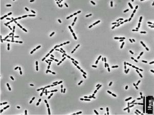
A diverse group of microbes live within the digestive systems of ruminants. Ruminants include cloven-hoofed animals with plant-based diets such as cows, sheep, deer, and goats that degrade plant materials in a specialized foregut organ, the rumen (Leshine SB). The microbes allow the animals to break down complex plant materials such as amino acids, cellulose, starch, and sugars into simpler products that can then be broken down by the animal’s own metabolism. Ruminants have a four-chambered gut, and these microorganisms live primarily in the rumen. One particularly important bacterial genus that takes part in the degradation of cellulose is Ruminococcus. Ruminococcus bacteria break down the plant fiber into the monosaccharide glucose, which can then be further broken down through glycolysis. This symbiotic relationship enables ruminants to digest this fiber without having to encode for more enzymes in their own genomes to do this job. The relationship with microbes provides ruminants with about 15% of their caloric intake. The Ruminococcus genus, which includes Ruminococcus albus and Ruminococcus flavifaciens, is just one of many microbes living in the rumen. Others include Megasphaera, Fibrobacter, Streptococcus, Escherichia, Chytridiomycetes fungi, and methanogens. It is predicted that 70% of microbes in the rumen have yet to be identified (Cho SJ). The proportions of microbes present vary greatly depending upon the diet of the ruminant. For this reason, the diets of the animals greatly impact their own health, and also effects consumers of their meat and the environment as a whole.
The Four Chambered Stomach and Function of Ruminant Microbes
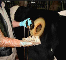
Ruminants have stomachs with four chambers; the rumen, the reticulum, the omasum, and the abomasums. Food is first mixed with saliva and passed to the rumen, where it is mechanically broken down to smaller pieces and then passed to the reticulum. Here the food is further broken down and also separated from indigestible non-food items before it is formed into cuds. These cuds are then regurgitated into the animal’s mouth, where it is re-chewed, and then re-swallowed back into the rumen. The majority of the anaerobic microbes that aid in cellulose breakdown inhabit the rumen, and during this step of digestion, fermentation begins. The partially digested food then moves to the omasum, where water, vitamins, and short chain fatty-acids from fermentation are absorbed into the animal’s body. pH is decreased and enzymes are released to further break down the material, before it is passed to the abomasums. The abomasums is the equivalent of the human stomach; material is further broken down and then passed to the small and large intestines where nutrients are absorbed before the waste is excreted (Leschine, SB). This process takes about 9-12 hours (Leshine, SB).
The rumen is an especially important chamber of the stomach, because it is here that cellulose is broken down into glucose, a sugar that can be used by the body. It is full of microbes that appear as soon as 38 hours after the animal’s birth (McAllister, TA). Without the microbes, the ruminants would get about 15% less nutrients from their food. The ruminal bacteria themselves are also digested by the animals in their small intestines, and further serve as a source of nutrients (Leschine SB). There are hundreds of different kinds of bacteria in the rumen which aid in this process, 85-95% which have yet to be cultured and 70% which have not yet been identified (Cho SJ). F. succinogenes, R. albus, and R. flavefaciends are the principle cellulose degrading oganisms (Leschine SB).
However, within the different areas of the rumen the types of microbes differ. Using PCR to amplify 16s rDNA sequences of microbes in the rumen, the fluid, solid, and epithelium were determined to host different populations (Cho SJ). The fluid had high levels of Cytophaga-Flexibacter-Bacteroides (67.5%), low G+C gram-positive bacteria (30%), and 2.5% proteobacteria. The solid was found to have the highest levels of low G+C gram positive bacteria (75.7%), 10.8% CFB, 5.4% proteobacteria and high G+C gram positive bacteria, and 2.7% spirochaetes. The epithelium had 94% CFB and 5.6% low G+C gram positive bacteria (Cho SJ). This suggests that these microbes play specific roles in rumen function. The most abundant organisms can effectively degrade and colonize the plant material, and can utilize a wide range of substrates, thus outcompeting the other microbes.
Within the rumen, it is necessary for the microbes to be able to attach to the plant material. Fungi disrupt the exterior recalcitrant tissue of the feed creating holes to make it more susceptible to enzymes (Weimer PJ). Primary colonizing microbes then create glycocalyx-enclosed microcolonies, which are joined by secondary microbes from the ruminal fluid, forming the multispecies biofilm (McAllister). This biofilm produces enzymes needed for the breakdown of cellulose. Using a complex metabolic pathway, these microbes turn cellulose into materials that can be used as energy sources by the ruminant animal as well as the microbe community.
Metabolic Pathway
Include some current research in each topic, with at least one figure showing data.
Factors that Effect the Microbe Population
Include some current research in each topic, with at least one figure showing data.
picuture will be graph showing rumen pH/bacterial contents
Conclusion
Overall paper length should be 3,000 words, with at least 3 figures.
References
Slonczewski JL and Foster JW. 2009. Microbiology: An Evolving Science. 2nd Edition. W.W. Norton, New York: 822-825.
Edited by KatiePruett, a student of Nora Sullivan in BIOL187S (Microbial Life) in The Keck Science Department of the Claremont Colleges Spring 2013.


