Change in the Pharyngeal Microbial Environment after a Tonsillectomy
By Lindsey Abramson
Introduction
Tonsillectomies, the surgical removal of the tonsils, are performed on individuals with chronic tonsillitis infections, recurrent strep throat, enlarged tonsils, and other issues such as cancerous cells (Brietzke and Andreoli, 2021). The goal of this radical procedure is the eradication of bacteria assuming antibiotics have been frequently used and failed / resistance was developed (Yildizoglu et al., 2015). Streptococcus pyogenes (also called group A streptococci, or GAS) is the causative agent bacterium of strep throat, one of the leading causes that push individuals towards an elective tonsillectomy (Slonczewski et al., 2024). Although viruses are also common, other problematic bacteria include: Staphylococcus aureus, Haemophilus influenzae, Streptococcus pneumoniae, Escherichia coli, and Pseudomonas aeruginosa (Yildizoglu et al., 2015).

At right is a sample image insertion. It works for any image uploaded anywhere to MicrobeWiki.
The insertion code consists of:
Double brackets: [[
Filename: PHIL_1181_lores.jpg
Thumbnail status: |thumb|
Pixel size: |300px|
Placement on page: |right|
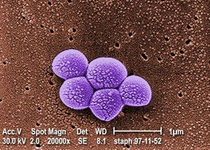
Legend/credit: Magnified 20,000X, this colorized scanning electron micrograph (SEM) depicts a grouping of methicillin resistant Staphylococcus aureus (MRSA) bacteria. Photo credit: CDC. Every image requires a link to the source.
Closed double brackets: ]]
Other examples:
Bold
Italic
Subscript: H2O
Superscript: Fe3+
Sample citations: [1]
[2]
A citation code consists of a hyperlinked reference within "ref" begin and end codes.
To repeat the citation for other statements, the reference needs to have a names: "<ref name=aa>"
The repeated citation works like this, with a forward slash.[1]
Section 1
Include some current research, with at least one figure showing data.
Every point of information REQUIRES CITATION using the citation tool shown above.
Section 2
Include some current research, with at least one figure showing data.
Section 3
Include some current research, with at least one figure showing data.
Section 4
Conclusion
References
Authored for BIOL 238 Microbiology, taught by Joan Slonczewski,at Kenyon College,2024
