Ehrlichia: tick-borne pathogen in canines and humans: Difference between revisions
| Line 13: | Line 13: | ||
<br><i>Italic</i> | <br><i>Italic</i> | ||
<br><b>Subscript:</b> H<sub>2</sub>O | <br><b>Subscript:</b> H<sub>2</sub>O | ||
<br><b>Superscript:</b | <br><b>Superscript:</b> | ||
Ehrlichia is an obligate intracellular bacterium of the family Anaplasmataceae- a classified α-proteobacteria. These Gram- negative coccobacilli, are best known for their etiological agency in the transmission of a group of tick-borne illnesses known as Ehrlichiosis (Ismail et al. 2011). All members of the genus Ehrlichia are pathogenic, causing mild to severe symptoms, specifically in humans and canines. This genus includes E.chaffeensis, E.ewingii, and E.canis (Nicholson et al. 2010). These bacteria are considered the agents responsible for the emergence of human monocytotropic ehrlichiosis and canine monocytotropic ehrlichiosis. The Ehrlichial agents are hosted in vertebrate reservoirs and transmitted to both dogs and humans by “hard” tick vectors. Like many other tick-borne illnesses the demographic distribution of Ehrlichiosis is greatest in regions with the highest density of primary tick vector populations; namely the Lone Star Tick (Amblyomma americanum) and the Brown Dog Tick (Rhipicephalus sanguineus) (Nicholson et al 2010). | Ehrlichia is an obligate intracellular bacterium of the family Anaplasmataceae- a classified α-proteobacteria. These Gram- negative coccobacilli, are best known for their etiological agency in the transmission of a group of tick-borne illnesses known as Ehrlichiosis (Ismail et al. 2011). All members of the genus Ehrlichia are pathogenic, causing mild to severe symptoms, specifically in humans and canines. This genus includes E.chaffeensis, E.ewingii, and E.canis (Nicholson et al. 2010). These bacteria are considered the agents responsible for the emergence of human monocytotropic ehrlichiosis and canine monocytotropic ehrlichiosis. The Ehrlichial agents are hosted in vertebrate reservoirs and transmitted to both dogs and humans by “hard” tick vectors. Like many other tick-borne illnesses the demographic distribution of Ehrlichiosis is greatest in regions with the highest density of primary tick vector populations; namely the Lone Star Tick (Amblyomma americanum) and the Brown Dog Tick (Rhipicephalus sanguineus) (Nicholson et al 2010). | ||
Globally, the two most common forms of disease causing Erlichia are Ehrlichia canis and Ehrlichia chaffeensis. The first case of Ehrlichiosis emerged from Africa in the mid 1930’s, reported by Donatien and Lestoquard. This canine monocytic ehrlichiosis did not gain meaningful attention until the 1970s when a significant number of cases were reported in German shepherds during the Vietnam War (Harrus et.al 2010). The disease is composed of 3 distinct phases described as acute, subclinical, and chronic. Symptoms are similar for all stages however the chronic stage is exceedingly more severe than the acute and subclinical stages. Dogs entering the chronic phase of the disease must undergo intense and expensive treatment and in most cases have little hope in terms of survival. E.canis has been reported to infect all breeds of dog; however the German shepherd seems to be most susceptible breed having the highest mortality rate compared to other dogs infected with the bacteria (Harrus et.al 2010). The tick vector responsible for transmission of Ehrlichiosis in canines is the brown dog tick. Until recently E.canis was thought to only infect canines. However, in 2006 a study was done that isolated E.canis in human patients proving compatibility for human pathogenic infection in addition to E.chaffeensis and E.ewingii (Nicholson et.al 2010). | Globally, the two most common forms of disease causing Erlichia are Ehrlichia canis and Ehrlichia chaffeensis. The first case of Ehrlichiosis emerged from Africa in the mid 1930’s, reported by Donatien and Lestoquard. This canine monocytic ehrlichiosis did not gain meaningful attention until the 1970s when a significant number of cases were reported in German shepherds during the Vietnam War (Harrus et.al 2010). The disease is composed of 3 distinct phases described as acute, subclinical, and chronic. Symptoms are similar for all stages however the chronic stage is exceedingly more severe than the acute and subclinical stages. Dogs entering the chronic phase of the disease must undergo intense and expensive treatment and in most cases have little hope in terms of survival. E.canis has been reported to infect all breeds of dog; however the German shepherd seems to be most susceptible breed having the highest mortality rate compared to other dogs infected with the bacteria (Harrus et.al 2010). The tick vector responsible for transmission of Ehrlichiosis in canines is the brown dog tick. Until recently E.canis was thought to only infect canines. However, in 2006 a study was done that isolated E.canis in human patients proving compatibility for human pathogenic infection in addition to E.chaffeensis and E.ewingii (Nicholson et.al 2010). | ||
Revision as of 22:16, 24 April 2013
Introduction

At right is a sample image insertion. It works for any image uploaded anywhere to MicrobeWiki. The insertion code consists of:
Double brackets: [[
Filename: PHIL_1181_lores.jpg
Thumbnail status: |thumb|
Pixel size: |300px|
Placement on page: |right|
Legend/credit: Electron micrograph of the Ebola Zaire virus. This was the first photo ever taken of the virus, on 10/13/1976. By Dr. F.A. Murphy, now at U.C. Davis, then at the CDC.
Closed double brackets: ]]
Other examples:
Bold
Italic
Subscript: H2O
Superscript:
Ehrlichia is an obligate intracellular bacterium of the family Anaplasmataceae- a classified α-proteobacteria. These Gram- negative coccobacilli, are best known for their etiological agency in the transmission of a group of tick-borne illnesses known as Ehrlichiosis (Ismail et al. 2011). All members of the genus Ehrlichia are pathogenic, causing mild to severe symptoms, specifically in humans and canines. This genus includes E.chaffeensis, E.ewingii, and E.canis (Nicholson et al. 2010). These bacteria are considered the agents responsible for the emergence of human monocytotropic ehrlichiosis and canine monocytotropic ehrlichiosis. The Ehrlichial agents are hosted in vertebrate reservoirs and transmitted to both dogs and humans by “hard” tick vectors. Like many other tick-borne illnesses the demographic distribution of Ehrlichiosis is greatest in regions with the highest density of primary tick vector populations; namely the Lone Star Tick (Amblyomma americanum) and the Brown Dog Tick (Rhipicephalus sanguineus) (Nicholson et al 2010).
Globally, the two most common forms of disease causing Erlichia are Ehrlichia canis and Ehrlichia chaffeensis. The first case of Ehrlichiosis emerged from Africa in the mid 1930’s, reported by Donatien and Lestoquard. This canine monocytic ehrlichiosis did not gain meaningful attention until the 1970s when a significant number of cases were reported in German shepherds during the Vietnam War (Harrus et.al 2010). The disease is composed of 3 distinct phases described as acute, subclinical, and chronic. Symptoms are similar for all stages however the chronic stage is exceedingly more severe than the acute and subclinical stages. Dogs entering the chronic phase of the disease must undergo intense and expensive treatment and in most cases have little hope in terms of survival. E.canis has been reported to infect all breeds of dog; however the German shepherd seems to be most susceptible breed having the highest mortality rate compared to other dogs infected with the bacteria (Harrus et.al 2010). The tick vector responsible for transmission of Ehrlichiosis in canines is the brown dog tick. Until recently E.canis was thought to only infect canines. However, in 2006 a study was done that isolated E.canis in human patients proving compatibility for human pathogenic infection in addition to E.chaffeensis and E.ewingii (Nicholson et.al 2010).
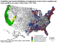
The primary agent for Human Monocytic Ehrlichiosis is E. chaffeensis, transmitted by the Lone Star tick. Ehrlichiosis in human patients was first seen in the United States in 1986 and has since emerged as one of the leading tick-borne illness in the United States, next to Rocky Mounted Spotted fever and Lyme’s Disease (Ismail et al 2010). E.ewingii has also emerged in recent years as the causative agent of a more mild case of human ehrlichiosis. Most cases of E. ewingii since 1999 have been reported in patients with HIV or other infections that coincide with decreased immune response (Ismail et al. 2011). Most occurrences of infection by E.canis and E.chaffeensis are predominantly reported in the south central and south eastern United States as this is the natural ecological range of the known tick vectors.
Section 1
Include some current research in each topic, with at least one figure showing data.

Section 2
Include some current research in each topic, with at least one figure showing data.

Section 3
Include some current research in each topic, with at least one figure showing data.
Conclusion
Overall paper length should be 3,000 words, with at least 3 figures.
References
Edited by student of Joan Slonczewski for BIOL 238 Microbiology, 2009, Kenyon College.
