File:02-0485 1b.jpg: Difference between revisions
From MicrobeWiki, the student-edited microbiology resource
(a) Hematoxylin and eosin stain of a lesion specimen showing definitive Buruli ulcer disease in the preulcerative stage (original magnification 50x). Notice the psoriasiform epidermal hyperplasia (H), superficial dermal lichenoid inflammatory infiltrate (I) |
No edit summary |
||
| Line 1: | Line 1: | ||
a) Hematoxylin and eosin stain of a lesion specimen showing definitive Buruli ulcer disease in the preulcerative stage (original magnification 50x). Notice the psoriasiform epidermal hyperplasia (H), superficial dermal lichenoid inflammatory infiltrate (I), and necrosis of subcutaneous tissues (N). B) Ziehl-Neelsen stain of the same nodule, showing abundant colonies of acid-fast bacilli in the necrotic subcutaneous tissues (original magnification 100x). | a) Hematoxylin and eosin stain of a lesion specimen showing definitive Buruli ulcer disease in the preulcerative stage (original magnification 50x). Notice the psoriasiform epidermal hyperplasia (H), superficial dermal lichenoid inflammatory infiltrate (I), and necrosis of subcutaneous tissues (N). B) Ziehl-Neelsen stain of the same nodule, showing abundant colonies of acid-fast bacilli in the necrotic subcutaneous tissues (original magnification 100x). | ||
From the CDC | |||
Latest revision as of 01:59, 27 August 2009
a) Hematoxylin and eosin stain of a lesion specimen showing definitive Buruli ulcer disease in the preulcerative stage (original magnification 50x). Notice the psoriasiform epidermal hyperplasia (H), superficial dermal lichenoid inflammatory infiltrate (I), and necrosis of subcutaneous tissues (N). B) Ziehl-Neelsen stain of the same nodule, showing abundant colonies of acid-fast bacilli in the necrotic subcutaneous tissues (original magnification 100x).
From the CDC
File history
Click on a date/time to view the file as it appeared at that time.
| Date/Time | Thumbnail | Dimensions | User | Comment | |
|---|---|---|---|---|---|
| current | 00:30, 27 August 2009 | 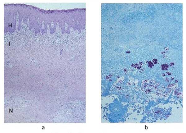 | 600 × 428 (58 KB) | Brlim (talk | contribs) | a) Hematoxylin and eosin stain of a lesion specimen showing definitive Buruli ulcer disease in the preulcerative stage (original magnification 50x). Notice the psoriasiform epidermal hyperplasia (H), superficial dermal lichenoid inflammatory infiltrate (I |
You cannot overwrite this file.
File usage
The following page uses this file:
