File:10247 lores.jpg: Difference between revisions
From MicrobeWiki, the student-edited microbiology resource
(This scanning electron micrograph (SEM) depicted three views of a single Gram-negative Neisseria gonorrhoeae bacterium. Under this highly-magnified view, the “roughened” texture of the bacterium’s cell wall is made visible. As a Gram-negative bacter) |
(No difference)
|
Latest revision as of 08:07, 28 August 2009
This scanning electron micrograph (SEM) depicted three views of a single Gram-negative Neisseria gonorrhoeae bacterium. Under this highly-magnified view, the “roughened” texture of the bacterium’s cell wall is made visible. As a Gram-negative bacterium, N. gonorrhoeae possess a thinner cell wall than its Gram-positive cousins, composed of peptidoglycan molecular layers that are sandwiched between a lipid membrane layer. Courtesy of CDC.
File history
Click on a date/time to view the file as it appeared at that time.
| Date/Time | Thumbnail | Dimensions | User | Comment | |
|---|---|---|---|---|---|
| current | 08:07, 28 August 2009 | 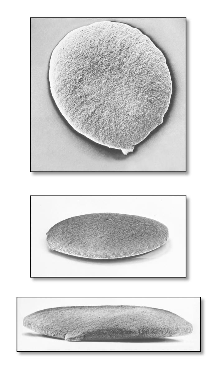 | 700 × 1,213 (90 KB) | Mhigashi (talk | contribs) | This scanning electron micrograph (SEM) depicted three views of a single Gram-negative Neisseria gonorrhoeae bacterium. Under this highly-magnified view, the “roughened” texture of the bacterium’s cell wall is made visible. As a Gram-negative bacter |
You cannot overwrite this file.
File usage
The following page uses this file:
