File:Filamentous phage.jpg: Difference between revisions
From MicrobeWiki, the student-edited microbiology resource
(Filamentous phage(s) under an electron microscope (left), and artist's rendition, and some computer-generated models of varying proteins found in the head, neck, and tail fibers.) |
|||
| Line 1: | Line 1: | ||
== Summary == | == Summary == | ||
Filamentous phage(s) under an electron microscope (left), | Filamentous phage(s) under an electron microscope (left), an artist's rendition, and some computer-generated models of varying proteins found in the head, neck, and tail fibers. | ||
Latest revision as of 02:37, 11 December 2020
Summary
Filamentous phage(s) under an electron microscope (left), an artist's rendition, and some computer-generated models of varying proteins found in the head, neck, and tail fibers.
File history
Click on a date/time to view the file as it appeared at that time.
| Date/Time | Thumbnail | Dimensions | User | Comment | |
|---|---|---|---|---|---|
| current | 02:23, 11 December 2020 | 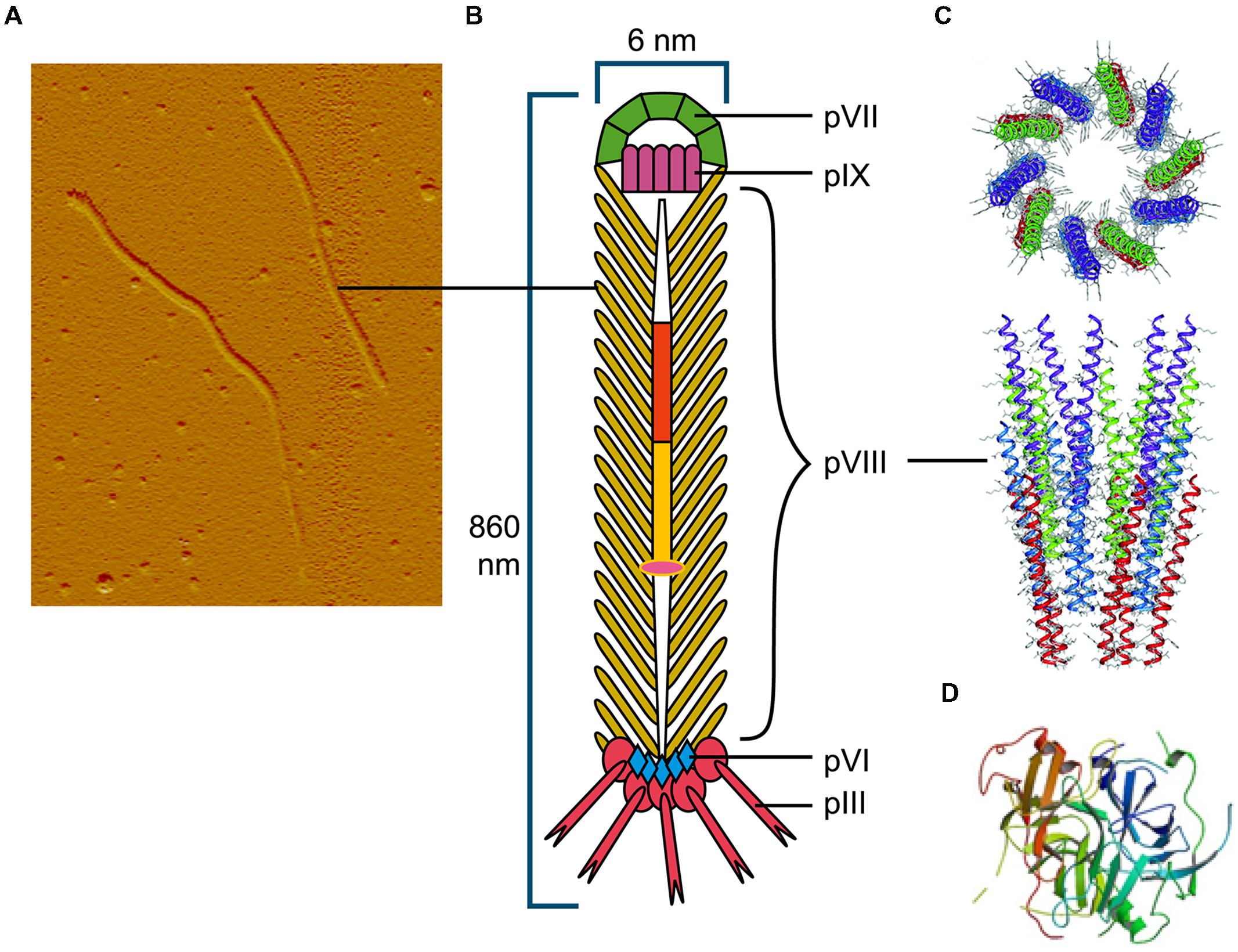 | 2,133 × 1,640 (392 KB) | Cerny1 (talk | contribs) | Filamentous phage(s) under an electron microscope (left), and artist's rendition, and some computer-generated models of varying proteins found in the head, neck, and tail fibers. |
You cannot overwrite this file.
File usage
The following page uses this file:
