File:Fullygrownparasite.png
From MicrobeWiki, the student-edited microbiology resource

No higher resolution available.
Fullygrownparasite.png (153 × 442 pixels, file size: 29 KB, MIME type: image/png)
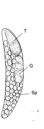
Figure 1 shows a fully grown parasite which would live inside its host. (T) points to the trophocyte, (G) points to the gonocyte, and (Sp) shows the sporocytes. (Image credited to froniters in microbiology, https://www.ncbi.nlm.nih.gov/pmc/articles/PMC3428600/figure/F1/ )
File history
Click on a date/time to view the file as it appeared at that time.
| Date/Time | Thumbnail | Dimensions | User | Comment | |
|---|---|---|---|---|---|
| current | 15:20, 19 April 2018 | 153 × 442 (29 KB) | Corhoski (talk | contribs) | Figure 1 shows a fully grown parasite which would live inside its host. (T) points to the trophocyte, (G) points to the gonocyte, and (Sp) shows the sporocytes. (Image credited to frontiers in microbiology, https://www.ncbi.nlm.nih.gov/pmc/articles/PMC... | |
| 15:16, 19 April 2018 | 153 × 442 (29 KB) | Corhoski (talk | contribs) | Figure 1 shows a fully grown parasite which would live inside its host. (T) points to the trophocyte, (G) points to the gonocyte, and (Sp) shows the sporocytes. (Image credited to froniters in microbiology, https://www.ncbi.nlm.nih.gov/pmc/articles/PMC... |
You cannot overwrite this file.
File usage
The following page uses this file:
