Human Chromosomal Integration of Latent State Human Herpes Virus 6 (HHV-6): Difference between revisions
From MicrobeWiki, the student-edited microbiology resource
| Line 25: | Line 25: | ||
==Latency through Human Chromosomal Integration== | ==Latency through Human Chromosomal Integration== | ||
[[Image:Integration.jpg|thumb|400px|right| [http://www.google.com/imgres?imgurl=http://schaechter.asmblog.org/.a/6a00d8341c5e1453ef0134860d4c84970c-350wi&imgrefurl=http://schaechter.asmblog.org/schaechter/2010/08/when-the-end-is-the-story.html&usg=__JTT4HmjU8tYFoKnz9jpyvS2avUw=&h=256&w=350&sz=11&hl=en&start=0&zoom=1&tbnid=FobJXAZiuuliUM:&tbnh=132&tbnw=181&prev=/images%3Fq%3DHHV-6%2Bchromosomal%2Bintegration%26um%3D1%26hl%3Den%26sa%3DN%26biw%3D1280%26bih%3D837%26tbs%3Disch:1&um=1&itbs=1&iact=rc&dur=404&ei=MmvPTO6kCYOGswbOsoH1AQ&oei=MmvPTO6kCYOGswbOsoH1AQ&esq=1&page=1&ndsp=27&ved=1t:429,r:7,s:0&tx=115&ty=85 Figure 5.]FISH visualization of HHV-6 integration into the telomere region of chromosome 9 in metaphase and interphase cells.] | [[Image:Integration.jpg|thumb|400px|right| [http://www.google.com/imgres?imgurl=http://schaechter.asmblog.org/.a/6a00d8341c5e1453ef0134860d4c84970c-350wi&imgrefurl=http://schaechter.asmblog.org/schaechter/2010/08/when-the-end-is-the-story.html&usg=__JTT4HmjU8tYFoKnz9jpyvS2avUw=&h=256&w=350&sz=11&hl=en&start=0&zoom=1&tbnid=FobJXAZiuuliUM:&tbnh=132&tbnw=181&prev=/images%3Fq%3DHHV-6%2Bchromosomal%2Bintegration%26um%3D1%26hl%3Den%26sa%3DN%26biw%3D1280%26bih%3D837%26tbs%3Disch:1&um=1&itbs=1&iact=rc&dur=404&ei=MmvPTO6kCYOGswbOsoH1AQ&oei=MmvPTO6kCYOGswbOsoH1AQ&esq=1&page=1&ndsp=27&ved=1t:429,r:7,s:0&tx=115&ty=85 Figure 5.]FISH visualization of HHV-6 integration into the telomere region of chromosome 9 in metaphase and interphase cells.] | ||
Revision as of 01:57, 2 November 2010
By: Kerri-Lynn Conrad
Introduction

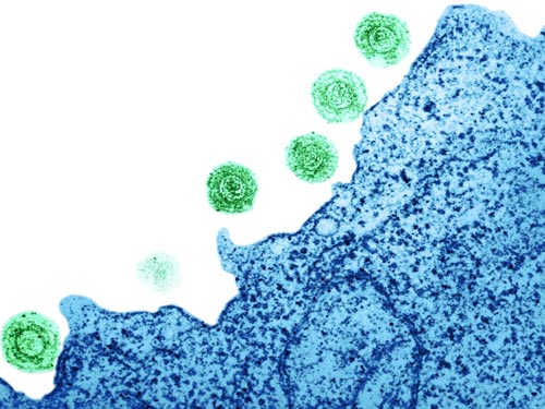
Figure 1. Transmission electron micrograph visualization of Human Herpes Virus-6 (HHV-6) on the surface of a human lymphocyte. HHV-6 is the causative agent of roseola, a disease that effects nearly every human infant.
Genomic Structure of HHV-6

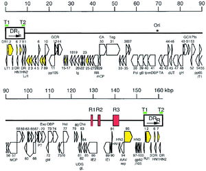
Figure 2. Genomic organization of HHV-6B. The asterisk indicates the start of lytic genomic replication. Viral telomeric sequences are indicated by green bars T1 and T2. Open arrows represent protein coding regions.
HHV-6 Replication Cycle

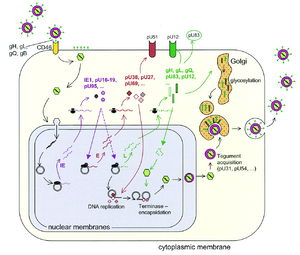
Figure 3. Lytic replication cycle of HHV-6.
Epidemiology
Pathophysiology of HHV-6 Infection

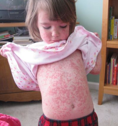
Figure 4. HHV-6 infection results in roseola, often diagnosed by a characteristic red rash. The roseola rash can form as either raised bumps or can be flat. It is non-contagious and is usually found on the neck, abdomen, back, and trunk, but can also be present on the arms and legs
Initial Infection
Latency in Healthy Children and Adults
Reactivation in Immunosuppressed Individuals
HHV-6 and Associated Disease States
Latency through Human Chromosomal Integration
[[Image:Integration.jpg|thumb|400px|right| Figure 5.FISH visualization of HHV-6 integration into the telomere region of chromosome 9 in metaphase and interphase cells.]
Transmission through Germ Line
Future Work
References
Edited by Kerri-Lynn Conrad, student of Joan Slonczewski for BIOL 375 Virology, 2010, Kenyon College.
