Leptospira Species in the Environment: Difference between revisions
From MicrobeWiki, the student-edited microbiology resource
No edit summary |
No edit summary |
||
| Line 13: | Line 13: | ||
<br><br>Other examples: | <br><br>Other examples: | ||
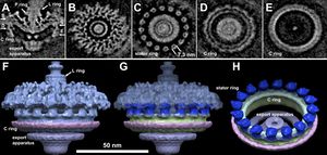
[[Image:Flagellamotility.jpg|thumg|300px|right|Figure 3.Molecular architecture of the intact flagellar motor of Leptospira spp.(A) Centered section parallel to the direction of the filament of an assymetric reconstruction of the Leptospiral motors. Panels B, C, D and E are horizontal cross sections. The locations of the sections are indicated in panel A. Panel B shows a section that lies inside the socket formed by the collar in periplasmic space. Section C transects the cytoplasmic side of the MS ring, just above the C ring. Sections D and E are located on the top and bottom of the C ring, respectively. Surface rendering of the Leptospira flagella motor is presented in panels F and G. ]] | [[Image:Flagellamotility.jpg|thumg|300px|right|Figure 3.Molecular architecture of the intact flagellar motor of Leptospira spp.(A) Centered section parallel to the direction of the filament of an assymetric reconstruction of the Leptospiral motors. Panels B, C, D and E are horizontal cross sections. The locations of the sections are indicated in panel A. Panel B shows a section that lies inside the socket formed by the collar in periplasmic space. Section C transects the cytoplasmic side of the MS ring, just above the C ring. Sections D and E are located on the top and bottom of the C ring, respectively. Surface rendering of the Leptospira flagella motor is presented in panels F and G.]] | ||
Revision as of 00:54, 23 April 2014
Introduction

Figure 2.The outer membrane (Om), inner membrane (IM), peptidoglycan layer (PG), and periplasmic flagellum (PF) in a 3-D reconstruction of intact L. interrogans (A) and L. biflexa (B).Zoom-in views reveal the detail of the cell envelope of L. interrogans (C) and L. biflexa (D). Panels E and F show the density profiles of L. interrogans and L. biflexa, respectively.
By Toni Miller
At right is a sample image insertion. It works for any image uploaded anywhere to MicrobeWiki. The insertion code consists of:
Double brackets: [[
Filename: LPS figure.gif
Thumbnail status: |thumb|
Pixel size: |300px|
Placement on page: |right|
Legend/credit: Electron micrograph of the Ebola Zaire virus. This was the first photo ever taken of the virus, on 10/13/1976. By Dr. F.A. Murphy, now at U.C. Davis, then at the CDC.
Closed double brackets: ]]
Other examples:
Introduce the topic of your paper. What microorganisms are of interest? Habitat? Applications for medicine and/or environment?
Section 1
Include some current research, with at least one figure showing data.


