Leptospira Species in the Environment: Difference between revisions
No edit summary |
No edit summary |
||
| Line 1: | Line 1: | ||
==Introduction== | ==Introduction== | ||
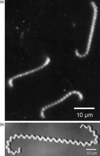
There are various species of Leptospira, all belonging to the spirochete phylum. The Leptospira bacteria are characterized by their unique shape. These Gram-negative bacteria are long and helical in shape, similar to other spirochetes, but contain hooked ends. The hooked ends occur as the result of the two periplasmic flagella located at either pole of the cell. [Fig. 1] These are responsible for the rapid dissemination of the organism into the desired host. [8]Leptospires are obligate aerobes and grow best at temperatures ranging from 28-30°C. [4] One of the most common species of Leptospira is Leptospira interrogans. This species is the most predominant pathogen of the Leptospira phylum and results in a disease called leptospirosis. Leptospirosis is a zoonotic disease predominant in the subtropical and tropical regions of the world. Most cases of leptospirosis in humans originate from a reservoir host, most commonly rodents, and are transmitted through water or contaminated soil. Specifically, L. interrogans colonizes the renal tubules of the host organism. [2] Leptospirosis in livestock can have detrimental effects on the agricultural industry, causing abortions, infertility and death. The effects of leptospirosis infection in humans can ranges from mild symptoms, such as a fever and a headache, to fatal problems causing death. These can include renal failure, jaundice, and pulmonary hemorrhage and are typically termed Weil's disease. [1,4] Contrarily, L. biflexa is a non pathogenic, saprophytic species that is incapable of infecting mammalian hosts. Although sharing many similarities in genetic and structural components, the L. biflexa lacks the genes necessary for virulence. The saprophytic species have been observed to have more genes however, serving as nutrient absorption. [2] With over 13 different species of pathogenic Leptospira and the recorded number of leptospirosis cases well over 1 million annually, Leptospira bacteria are ideal for research. Not only are there vast differences between these spirochetes and others, such as the bacteria responsible for Lyme Disease, but there are also vast differences between each species of Leptospira. [3] | |||
[[Image:Leptospira shape.jpg|thumb|200px|right|Figure 1. Dark field (a) and shadowed electron (b) photomicrographs of Leptospira spp.]] | [[Image:Leptospira shape.jpg|thumb|200px|right|Figure 1. Dark field (a) and shadowed electron (b) photomicrographs of Leptospira spp.]] | ||
Revision as of 22:56, 25 April 2014
Introduction
There are various species of Leptospira, all belonging to the spirochete phylum. The Leptospira bacteria are characterized by their unique shape. These Gram-negative bacteria are long and helical in shape, similar to other spirochetes, but contain hooked ends. The hooked ends occur as the result of the two periplasmic flagella located at either pole of the cell. [Fig. 1] These are responsible for the rapid dissemination of the organism into the desired host. [8]Leptospires are obligate aerobes and grow best at temperatures ranging from 28-30°C. [4] One of the most common species of Leptospira is Leptospira interrogans. This species is the most predominant pathogen of the Leptospira phylum and results in a disease called leptospirosis. Leptospirosis is a zoonotic disease predominant in the subtropical and tropical regions of the world. Most cases of leptospirosis in humans originate from a reservoir host, most commonly rodents, and are transmitted through water or contaminated soil. Specifically, L. interrogans colonizes the renal tubules of the host organism. [2] Leptospirosis in livestock can have detrimental effects on the agricultural industry, causing abortions, infertility and death. The effects of leptospirosis infection in humans can ranges from mild symptoms, such as a fever and a headache, to fatal problems causing death. These can include renal failure, jaundice, and pulmonary hemorrhage and are typically termed Weil's disease. [1,4] Contrarily, L. biflexa is a non pathogenic, saprophytic species that is incapable of infecting mammalian hosts. Although sharing many similarities in genetic and structural components, the L. biflexa lacks the genes necessary for virulence. The saprophytic species have been observed to have more genes however, serving as nutrient absorption. [2] With over 13 different species of pathogenic Leptospira and the recorded number of leptospirosis cases well over 1 million annually, Leptospira bacteria are ideal for research. Not only are there vast differences between these spirochetes and others, such as the bacteria responsible for Lyme Disease, but there are also vast differences between each species of Leptospira. [3]

By Toni Miller


Introduce the topic of your paper. What microorganisms are of interest? Habitat? Applications for medicine and/or environment?
Section 1
Include some current research, with at least one figure showing data.


