Myxoma Virus
Characteristics of the symbiont/pathogen
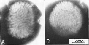
The myxoma genome is 161.8-kb long consisting of 171 unique genes (1F) making up this large, linear double-stranded DNA virus (2). The myxoma virus has a brick shaped viron and replicates only within the cytoplasm of the host cell (1).This enveloped virus is a member of the Poxvirus family and is of the Leporipoxvirus genus (3). The myxoma virus genome has terminal inverted repeats and covalently closed hairpin ends. The genes near the termini are not necessary for replication but are important in the virus’ virulence in infected animals (2).
Characteristics of the host
The host of the myxoma virus is European rabbits (Oryctolagus cuniculus). In this specific host, the myxoma virus causes a deadly infection called myxomatosis. The myxoma virus can infect other types of rabbits (including the American rabbit, Oryctolagus cuniculus), but only causes myxomatosis in European rabbits. The virus causes a tumor at the infection site of the rabbits and causes swelling of the adjacent connective tissues. Secondary myxoma tumors are found on other areas of infected rabbits (5).
Host-Symbiont Interaction
The myxoma virus is a parasitic symbiont to European rabbits. The relationship between the European rabbit and the myxoma virus is important to microbiologists because it is one of the few natural infections in which virulence can be equated with lethality (7). The myxoma virus causes lethal myxomatosis in European rabbits, a disease which causes tumor-like lesions, suppresses the immune system of the rabbits, and leads to a secondary gram negative bacterial infection. This relationship results in death of the European rabbit host (8).
Molecular Insights into the Symbiosis
The myxoma virus has a M-T7 protein that is specific in that it specifically binds rabbit IFN-gamma. By doing this, the M-T7 protein inhibits a regulatory cytokine that controls the host’s immune response to viral infections. This makes the M-T7 protein a critical virulence factor in the progression of myxomatosis (9). There are also several ways that poxviruses increase the virus' ability to survive in the wild. These mechanisms can involve the interference with major histocompatibility complex antigen presentation, inhibition of apoptosis, disruption of complement cascade, and inhibition of cytokines (2). Specifically, the myxoma virus uses inhibition of apoptosis to increase the virus' ability to survive in the wild. This inhibition of apoptosis is accomplished through the M11L genes which extend the host range and increases myxoma survival in the wild (9).
Ecological and Evolutionary Aspects
The myxoma virus origionated in North and South America as it evolved in close association with Sylvilagus (the cottontail rabbit). However, after 1950, the disease began to be used as a way to control rabbit populations in Europe, Australia, and Chile. Many different strands of the myxoma virus exist with virulances ranging from 50-95%. The more virulent strands were used as a way to control rabbit populations in these countries between 1950 and 1960 (10).
As of 2006, the myxoma virus is endemic of North America, South America, Europe, and Australia. In Brazil and Uruguay, the myxoma virus was found in rabbits of the Sylvilagus genus, however in other countries of South America, such a Brazil, the myxoma virus is found primarily in the European rabbit. In North America, the myxoma virus is found in rabbits of the Sylvilagus genus. On the other hand, in Australia and Europe, the myxoma virus is predominately found in European rabbits (O. cuniculus) (10).
Recent Discoveries

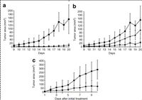
Current research is investigating the effect of the myxoma virus on cancerous tumors in mice. The myxoma virus was injected multiple times into cancerous tumors of mice. The viral injections resulted in a decreased size of the tumors (specifically B16F10 tumors). Additionally, systemic treatment of mice inhibited the development of lung metastasis. Mice were exposed to B16F10LacZ cells that cause lung metastasis and the mice that were treated with the myxoma virus did not develop observable tumors (11).
Other current research deals with the myxoma virus’ ability to kill most human malignant glioma cell lines. A single injection of the myxoma virus into the tumor dramatically increased the subject's median survival when compared to glioma tumors that were not treated with the myxoma virus. The median survival of myxoma treated subjects was 50.7 days compared to 47.3 days for the subjects that were not treated with myxoma virus. Additionally, 92% of the myxoma treated subjects were cured at the end of treatment and showed no remaining glioma tumors. This research also demonstrated that the myxoma virus is safe to administer to immunocompromised mice. It was found that the way in which the myxoma virus infected glioma cells was selective and this viral infection was long-lasting; lasting at least 42 days after initial infection (12).
References
Edited by Darcy Paulus, student of Grace Lim-Fong


