Physarum polycephalum: Difference between revisions
No edit summary |
No edit summary |
||
| Line 1: | Line 1: | ||
==Introduction== | ==Introduction== | ||
<i>Physarum polycephalum</i> is a species of acellular slime molds, or myxomycetes. Despite having a similar name to fungi, these organisms are protists. They are most commonly found in soil, moist vegetation, and the forest floor where it is cool, damp, and dark, where they contribute to the decomposition of dead vegetation. <i>P. polycephalum</i> has a very diverse life cycle, as they can range from being a microscopic single-celled amoeba, a multinucleated cell, or plasmodium, that can span several feet, morphing into a hardened mass known as a sclerotium, or forming small, delicate fruiting bodies that produce haploid spores. | |||
<br><br> | <br><br> | ||
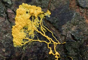
[[Image: | [[Image:1-plasmodial-slime-mould-nigel-downer.jpg|thumb|300px|right|Close-up of <i>P. polycephalum</i> creeping over a pine bark in a Norfolk pine forest. This single-celled organism with multiple nuclei forages for food by sending out branches (plasmodia) from a central location. Photograph taken by Nigel Downer on August 3rd, 2016.[https://fineartamerica.com/featured/1-plasmodial-slime-mould-nigel-downer.html].]] | ||
==Section 1 Genetics== | ==Section 1 Genetics== | ||
Revision as of 14:52, 9 December 2020
Introduction
Physarum polycephalum is a species of acellular slime molds, or myxomycetes. Despite having a similar name to fungi, these organisms are protists. They are most commonly found in soil, moist vegetation, and the forest floor where it is cool, damp, and dark, where they contribute to the decomposition of dead vegetation. P. polycephalum has a very diverse life cycle, as they can range from being a microscopic single-celled amoeba, a multinucleated cell, or plasmodium, that can span several feet, morphing into a hardened mass known as a sclerotium, or forming small, delicate fruiting bodies that produce haploid spores.

Section 1 Genetics
Include some current research, with at least one image.
Sample citations: [1]
[2]
A citation code consists of a hyperlinked reference within "ref" begin and end codes.
Section 2 Microbiome
Include some current research, with a second image.
Conclusion
Overall text length should be at least 1,000 words (before counting references), with at least 2 images. Include at least 5 references under Reference section.
References
- ↑ Hodgkin, J. and Partridge, F.A. "Caenorhabditis elegans meets microsporidia: the nematode killers from Paris." 2008. PLoS Biology 6:2634-2637.
- ↑ Bartlett et al.: Oncolytic viruses as therapeutic cancer vaccines. Molecular Cancer 2013 12:103.
- ↑ Lee G, Low RI, Amsterdam EA, Demaria AN, Huber PW, Mason DT. Hemodynamic effects of morphine and nalbuphine in acute myocardial infarction. Clinical Pharmacology & Therapeutics. 1981 May;29(5):576-81.
Edited by [Author Name], student of Joan Slonczewski for BIOL 116 Information in Living Systems, 2020, Kenyon College.
