Prevotella nigrescens

Prevotella species are a part of the oral flora. Prevotella nigrescens (P. nigrescens), a member of this family, was first discovered in within a group of bacteria termed Bacteroides intermedius.[1] Following this discovery, P. intermedia and P. nigrescens were found to play a role in the pathogenesis of periodontal and endodontic disease. More recently, P. nigrescens was also found to be associated with signs of carotid atherosclerosis even in patients without periodontitis and with endodontic infections.[2] [3]
As more information on the pathogenesis properties of P. nigrescens was uncovered, there was a demand to find a treatment. Under this pressure a potential alternative treatment, to antibiotics, for P. nigrescens was found in the use of bee glue.[4] Previous studies suggest that P. intermedia is more often found in periodontal sites whereas P. nigrescens is more frequently found in healthy gingivae. However, P. nigrescens have been implicated as a major periodontal pathogen. The site specificity of P. nigrescens remains unclear. [3] [4]
Cellular morphology and biochemistry

P. nigrescens are black-pigmented bacteroides that ferment carbohydrates. They are gram-negative, non-sporing, obligatory anaerobic, rod-shaped bacterium of the genus Prevotella that is commonly found in oral flora.
Cells grow in broth cultures are 0.3 to 0.7 µm wide by 1 to 2 µm. In agar, colonies are 0.5 to 2 mm in diameter, circular, low convex and smooth. In blood agar, the colonies are brown to black suggesting weakly hemolytic, and sometimes alpha-hemolytic, properties. Pigmentation is mainly towards the outer edges whereas the center remains creamy to dark brown. Strains grown on blood agar also exhibit red fluorescence under long-wave UV radiation (365nm).[1]
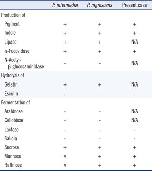
P. nigrescens uses mixed fermentation in anaerobic living conditions, breaking down dextrin, glucose, maltose, and sucrose. In the process of fermentation, they produce acetic, isobutryic, isovaleric, and succinic acids. Characterization using the API ZYM system revealed that P. nigrescens reacted with acid and alkaline phosphatases, phosphoamides, and β-glucosidases. The system showed weak reactions with butyrate esterase and caprylate esterase lipase. The principal respiratory quinones are unsaturated menaquinones. The G+C content of the DNA is 40 to 44 mol%.[1]
Differentiation from P. intermedia
Identification of samples as either P. intermedia or P. nigrescens can be made based on differences in malate and glutamate dehydrogenase electrophoretic mobility. Sodium dodecyl-polyacrylamide gel electrophoresis (SDS-PAGE) of whole cell samples show that P. nigrescens have 31 and 38 kDa proteins that are not present in P. intermedia.[5] Surface biotinylation of cells revealed that the 31 kDa protein is on the surface of the protein. Fimbria-like projections were also observed in negatively stained cells of P. nigrescens but not in P. intermedia.[5] Although no further research has been done on the 38 kDa protein, this protein may be of further help to distinguish both species as it stained strongly only in P. nigrescens. In terms of enzymatic differences, P. intermedia contain fast-moving enzymes whereas P. nigrescens have slower moving enzymes.[3] P. intermedia and P. nigrescens also differ in localization. P. nigrescens is almost three times more frequently isolated from infected root canals than P. intermedia.[3]

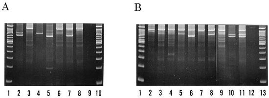
However, identifying the two strains using methods such as SDS-PAGE, restriction endonuclease analysis, ribotyping, and arbitrarily primer PCR (AP-PCR) are either time-consuming or have low levels of reproducibility. A newer method, PCR ribotyping, is expected to amplify the 16S and 23S spacer regions of bacterial rRNA genes by PCR. [6] It generates simple banding patterns that allows comparison between separate PCR amplifications. Since PCR ribotyping amplicons smaller than 600bp were poorly reproduced, higher discriminatory assays were run by using a combination of PCR ribotyping and AP-PCR fingerprinting. The combination assay identified seven types within the P. intermedia strain and ten types within the P. nigrescens strain, all of which were differentiable by a combination of their band patterns and DNA fingerprints.
Role in Disease
Periodontitis
P. nigrescens is part of the orange complex of subgingival and supragingival plaque. It is also strongly related to bacteria of the red complex, which is the most commonly used periodontal diagnosis and therefore is considered periodontal pathogens. [7] Much evidence supporting its pathogenic role comes from quantitative cultural studies, which is the comparison of certain pathogen levels in diseased sites to healthy sites.[8]
Recently, it was discovered that P. nigrescens is found more frequently in subgingival plaque of patients with periodontitis.[1] Periodontal sites are classified as either active, inactive or healthy using a number of widely accepted clinical criteria.[8] Researchers found a correlation between increased colonization of P. nigrescens and periodontitis.[1] Another group found that up to 70% of patients with localized and generalized forms of onset periodontitis had detectable levels of P. nigrescens in their subgingival plaque.[9] More recently, P. nigrescens was accepted as a putative periodontal pathogen for periodontal disease. Studies suggest that the root of its pathogenicity lies in that it seems to promote the release of inflammatory mediators.[10] [11]
Contradictory data showed that 9 of 21 subjects (15 periodontitis patients and 6 healthy volunteers) harbored more than one genotype of P. nigrescens. Several different types were found occupying different sites around the same tooth.[8] This data suggested that no specific relationship existed between disease status and colonization of same or different types of P. nigrescens.
Endodontic Infections
Carotid atherosclerosis
Treatment
The development of new therapies for treatment of oral cavity diseases is of great importance since the development of antibiotic resistance of certain microbes. One of these new treatments that have emerged is the use of propolis, more commonly known as bee glue.[4]
The biological activities of bee glue include antibacterial, antifungal, antiprotozoan, antiviral, antitumoral, immunomodulation, anti-inflammatory and other therapeutic properties.[4] It has been shown to be effective in treating gum infections due to P. nigrescens. Penicillin G, erythromycin, meropenem, metronidazole and tetracycline were found to inhibit 90% of the growth (MIC90) of P. intermedia and P. nigrescens strains at of 0.06, 0.13, 2.0, 4.0 and 4.0ug/mL respectively. iP. intermedia and P. nigrescens were found to be susceptible to three ethanolic extract of propolis at 128ug/mL.[4]
References
1. Shah HN, Gharbia SE (1992). Biochemical and chemical studies on strains designated Prevotella intermedia and proposal of a new pigmented species, Prevotella nigrescens sp. nov. Int J Syst Bacteriol 42: 542–6.
2. Yakob M., Söder B., Meurman J. H., Jogestrand T., Nowak J., Söder P.-Ö. (2011). Prevotella nigrescens and Porphyromonas gingivalis are associated with signs of carotid atherosclerosis in subjects with and without periodontitis. Journal of Periodontal Research 46 (6): 749–755.
3. Saheer E.G, Markus H, Haroun N.S, Anja K, Kari L, Michelle A. P., Deirdre A. D. (1994) Characterization of Prevotella intermedia and Prevotella nigrescens Isolates From Periodontic and Endodontic Infections. Journal of Periodontology 65(1): 56-61
4. Santos, F. A., Bastos, E. M. A., Rodrigues, P. H., de Uzeda, M., de Carvalho, M. A. R., de Macedo Farias, L., Moreira, E. S. A. (2002). Susceptibility of Prevotella intermedia/Prevotella nigrescens (and Porphyromonas gingivalis) to propolis (bee glue) and other antimicrobial agents. Anaerobe, 8(1), 9-15.
5. Fernández-Canigia L, Cejas D, Gutkind G, Radice M. (2015) Detection and genetic characterization of β-lactamases in Prevotella intermedia and Prevotella nigrescens isolated from oral cavity infections and peritonsillar abscesses. Anaerobe. 2015 Jan 23;33C:8-13.
6. Fukui K, Kato N, Kato H, Watanabe K, Tatematsu N (1999). Incidence of Prevotella intermedia and Prevotella nigrescens carriage among family members with subclinical periodontal disease. Journal of Clinical Microbiology 37 (10): 3141–5.
7. Stingu C.S., Schaumann R., Jentsch H., Eschrich K., Brosteanu O., Rodloff A.C (2013). Association of periodontitis with increased colonization by Prevotella nigrescens. Journal of Investigative and Clinical Dentistry 4 (1): 20–25.
8. Teanpaisan R, Douglas CW, Eley AR, Walsh TF (1996) Clonality of Porphyromonas gingivalis, Prevotella intermedia and Prevotella nigrescens isolated from periodontally diseased and healthy sites. Journal of Periodontal Research 31(6): 423-432
9. Mullally BH, Dace B, Shelburne CE, Wolff LF, Coulter WA. (2000) Prevalence of periodontal pathogens in localized and generalized forms of early-onset periodontitis. J Periodontal Res 35: 232–41.
10. Kikkert R, Laine ML, Aarden LA, van Winkelhoff AJ (2007). Activation of toll-like receptors 2 and 4 by Gram negative periodontal bacteria. Oral Microbiol Immunol 22: 145–51.
11. Airila-Mansson S, Soder B, Kari K, Meurman JH (2006). Influence of combinations of bacteria on the levels of prostaglandin E2, interleukin-1ß, and granulocyte elastase in gingival crevicular fluid and on the severity of periodontal disease. J Periodontol 77: 1025–31.
12. Kwang-Shik B, J. Craig B., Thomas R. S., Larry L. D. (2007) Occurrence of Prevotella nigrescens and Prevotella intermedia in infections of endodontic origin. Journal of Endodontics 23(10): 620-623
13. Yang JJ, Kwon TY, Seo MJ, Nam YS, Han CS, Lee HJ. (2013) 16S Ribosomal RNA Identification of Prevotella nigrescens from a Case of Cellulitis. Ann Lab Med 3(5):379-382.
Edited by Nancy Zhu, a student of Suzanne Kern in BIOL168L (Microbiology) in The Keck Science Department of the Claremont Colleges Spring 2015.
