File list
From MicrobeWiki, the student-edited microbiology resource
This special page shows all uploaded files.
| Date | Name | Thumbnail | Size | User | Description | Versions |
|---|---|---|---|---|---|---|
| 23:11, 12 June 2020 | Figure 11 373wiki.jpg (file) |  |
86 KB | Andreagomez-patron2020 | 1 | |
| 21:51, 12 June 2020 | Figure 10 373wiki.png (file) |  |
77 KB | Andreagomez-patron2020 | 1 | |
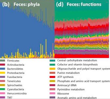
| 19:18, 12 June 2020 | Figure 9 373wiki.jpg (file) |  |
94 KB | Andreagomez-patron2020 | 1 | |
| 19:17, 12 June 2020 | Figure 8 373wiki.jpg (file) |  |
55 KB | Andreagomez-patron2020 | 1 | |
| 19:06, 12 June 2020 | Figure 7 373wiki.jpg (file) |  |
23 KB | Andreagomez-patron2020 | 1 | |
| 08:39, 12 June 2020 | Functionalredundancy.jpg (file) |  |
53 KB | Nhutran2021 | 1 | |
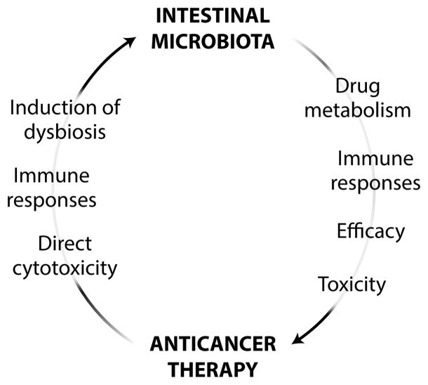
| 08:26, 12 June 2020 | Cancermicrobiome.jpg (file) |  |
39 KB | Nhutran2021 | 1 | |
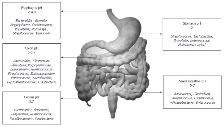
| 07:49, 12 June 2020 | Gut.jpg (file) |  |
77 KB | Nhutran2021 | 1 | |
| 04:29, 12 June 2020 | GSL Diversity.jpg (file) |  |
973 KB | Floydnichols2025 | 1 | |
| 04:23, 12 June 2020 | Salinity.jpg (file) |  |
1.76 MB | Floydnichols2025 | 1 | |
| 04:07, 12 June 2020 | Key Players.jpg (file) |  |
86 KB | Floydnichols2025 | 1 | |
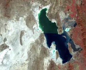
| 04:04, 12 June 2020 | Great Salt Lake.jpg (file) |  |
30 KB | Floydnichols2025 | 1 | |
| 03:52, 12 June 2020 | Fluid Inclusion.jpg (file) |  |
102 KB | Floydnichols2025 | 1 | |
| 02:55, 12 June 2020 | Boetius2.png (file) |  |
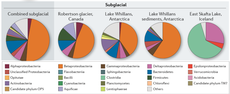
681 KB | Timothycoston2024 | Image displays bacterial diversity from a variety of subglacial environments. Figure modified from below source: Boetius, A., Anesio, A.M., Deming, J.W., Mikucki, J.A., and Rapp, J.Z., 2015, Microbial ecology of the cryosphere: sea ice and glacial habi... | 1 |
| 02:38, 12 June 2020 | Boetius.png (file) | 392 KB | Timothycoston2024 | Figure modified from below source: Boetius, A., Anesio, A.M., Deming, J.W., Mikucki, J.A., and Rapp, J.Z., 2015, Microbial ecology of the cryosphere: sea ice and glacial habitats: Nature Reviews, v. 13, p. 677-690. | 1 | |
| 17:51, 11 June 2020 | Desulforudis audaxviator.jpg (file) |  |
688 KB | Timothyellis2021 | 1 | |
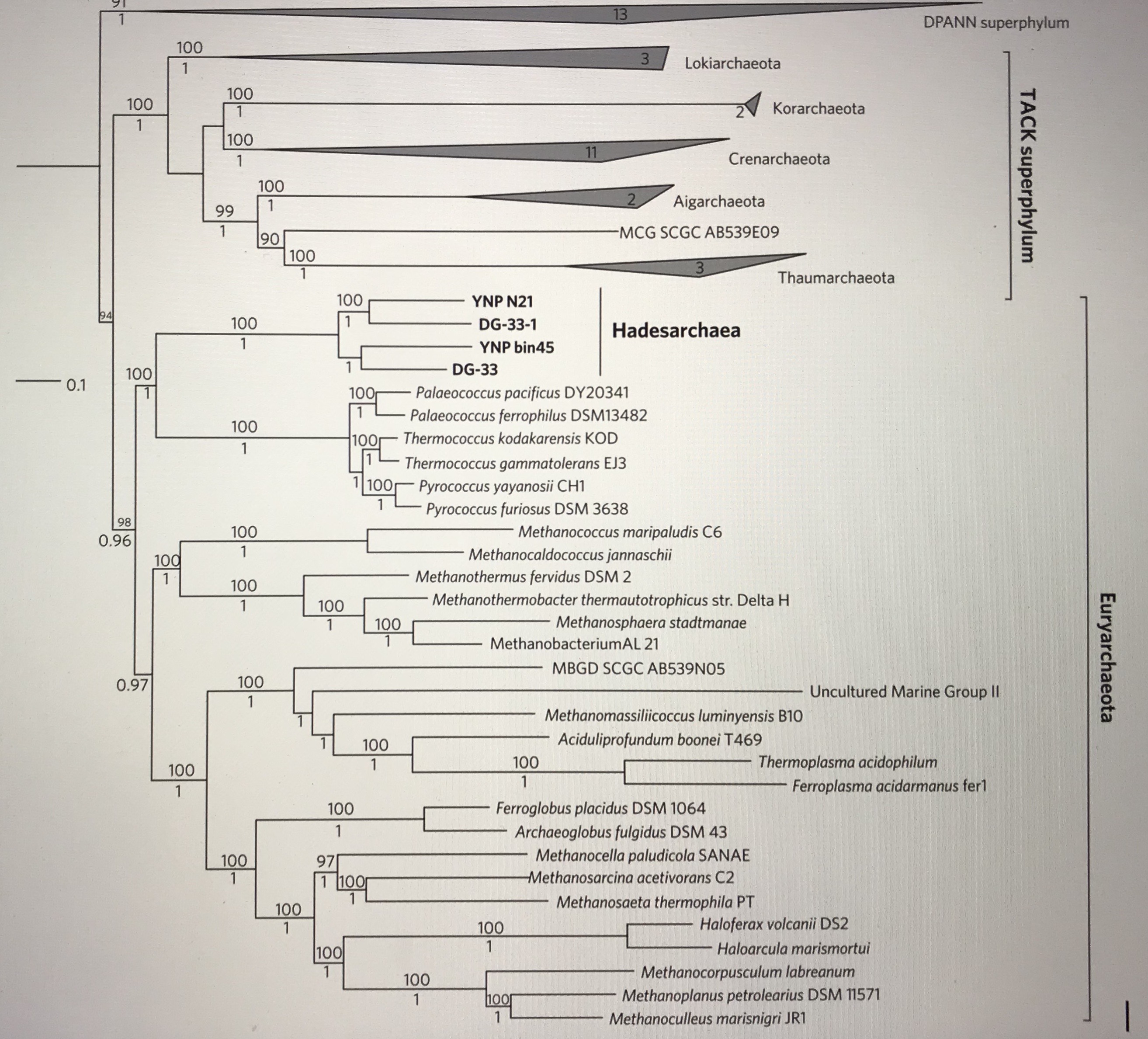
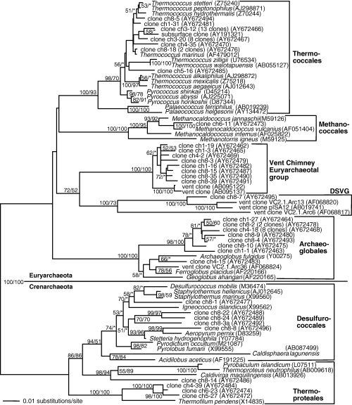
| 17:42, 11 June 2020 | Hadesarchaea-7128.JPG (file) |  |
1.15 MB | Timothyellis2021 | Phylogenetic tree | 1 |
| 16:22, 10 June 2020 | Atmdustsize.jpeg (file) |  |
57 KB | Robertgallo2021 | https://www.ncbi.nlm.nih.gov/pmc/articles/PMC2982008/ | 1 |
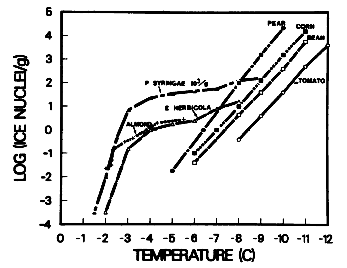
| 16:08, 10 June 2020 | Icenucleus.png (file) |  |
235 KB | Robertgallo2021 | Ice formation with microbe nuclei | 1 |
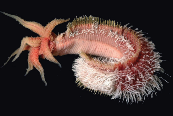
| 15:53, 10 June 2020 | Pompeiiworm.png (file) |  |
237 KB | Carolinewebster2021 | 1 | |
| 15:49, 10 June 2020 | Extremeyeast.jpeg (file) |  |
114 KB | Robertgallo2021 | Chart showing the survival of yeast at altitude from a study https://aem.asm.org/content/84/23/e01942-18#sec-2 | 1 |
| 15:02, 10 June 2020 | Atmlayers.png (file) |  |
375 KB | Robertgallo2021 | Layers and conditions of the atmosphere | 1 |
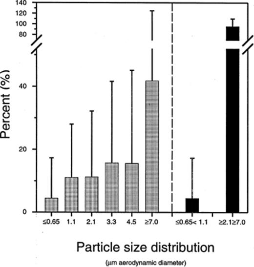
| 15:02, 10 June 2020 | Atmsizedistro.jpeg (file) |  |
29 KB | Robertgallo2021 | Distribution of atmospheric particle sizes | 1 |
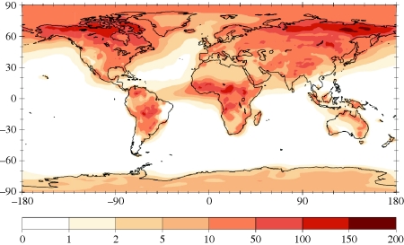
| 15:01, 10 June 2020 | Atmosphericmicrobemap.jpg (file) |  |
81 KB | Robertgallo2021 | Map of microbes in atmosphere from wind | 1 |
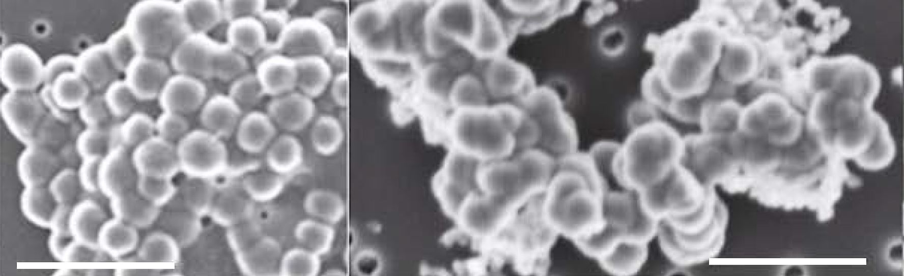
| 19:43, 6 June 2020 | SEM of cells and Si biomorphs from Dallol.jpg (file) |  |
109 KB | Rachelso2020 | Replaced with a larger image of the original. | 2 |
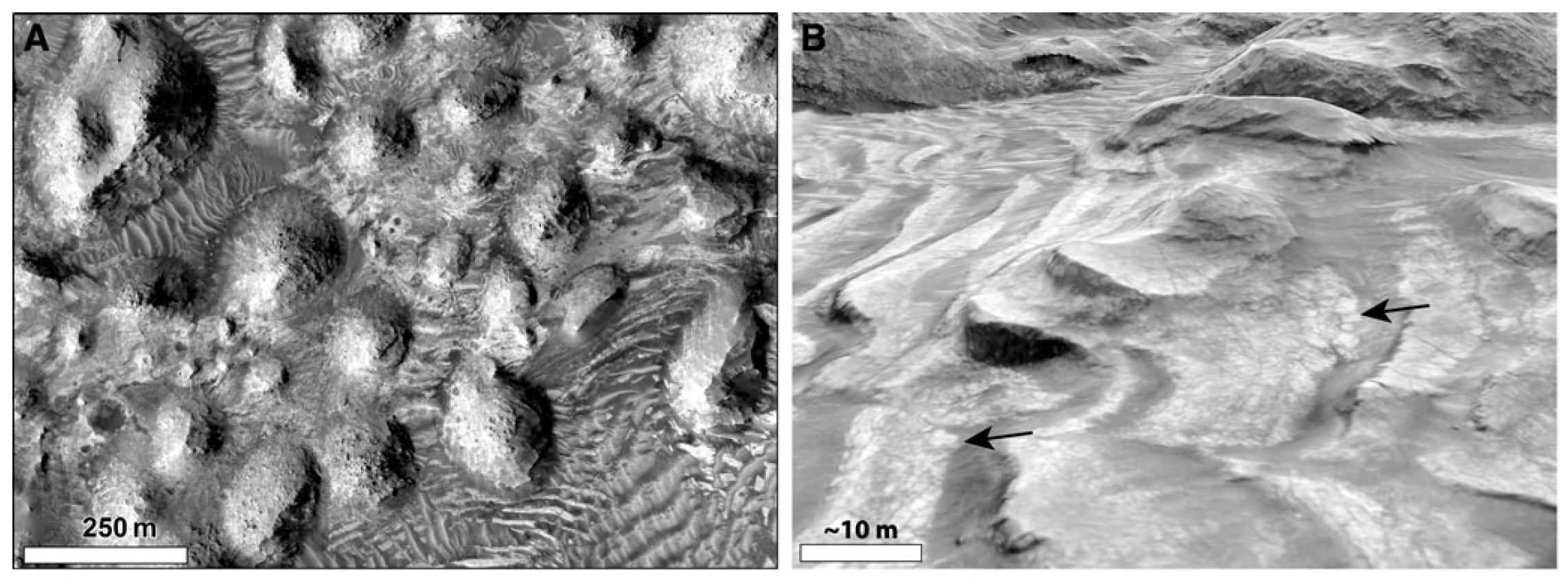
| 19:39, 6 June 2020 | Arabia Terra on Mars.JPG (file) |  |
309 KB | Rachelso2020 | Satellite images of Arabia Terra, a region on Mars. Source: https://www.liebertpub.com/doi/full/10.1089/ast.2018.1926 | 1 |
| 19:36, 6 June 2020 | SEM of mineral-encrusted ultrasmall cells from Dallol.JPG (file) |  |
155 KB | Rachelso2020 | Took out the bottom image because it was blurry. | 2 |
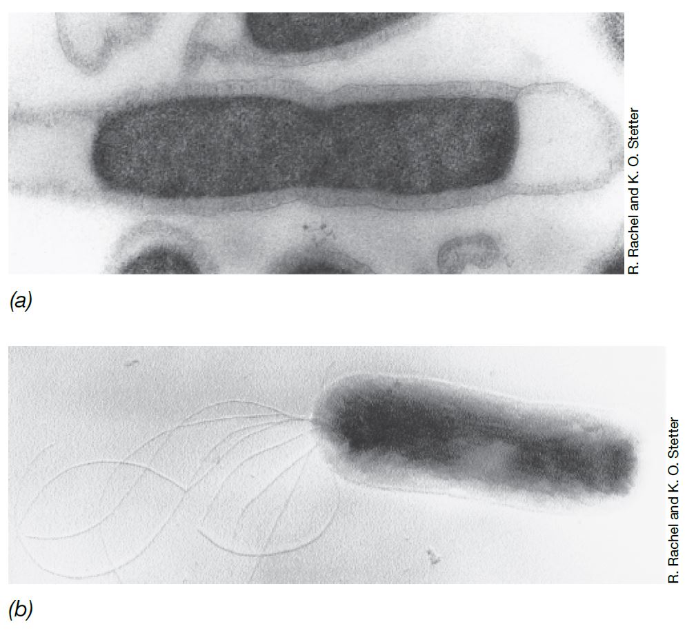
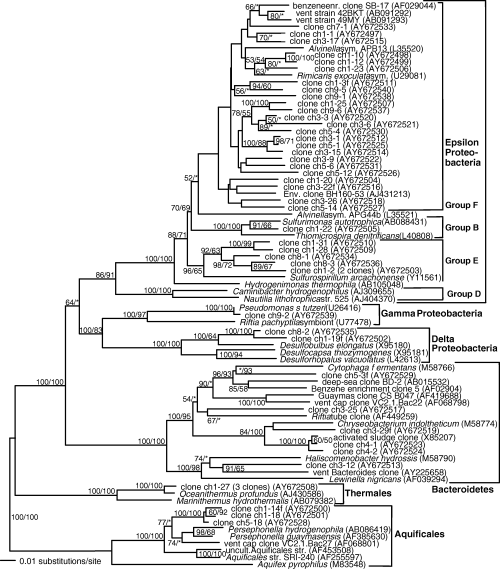
| 19:18, 6 June 2020 | Thermotoga and Aquifex.JPG (file) |  |
107 KB | Rachelso2020 | Electron micrographs of Thermotoga maritimus (top) and Aquifex pyrophilus (bottom). Source: Brock Biology of Microorganisms (15th edition) | 1 |
| 18:58, 6 June 2020 | San Francisco Bay Salt Ponds.jpg (file) |  |
744 KB | Rachelso2020 | High saline ponds in San Francisco Bay containing abundant Halobacteria. Source: https://commons.wikimedia.org/wiki/File:San_Francisco_Bay_Salt_Ponds.jpg | 1 |
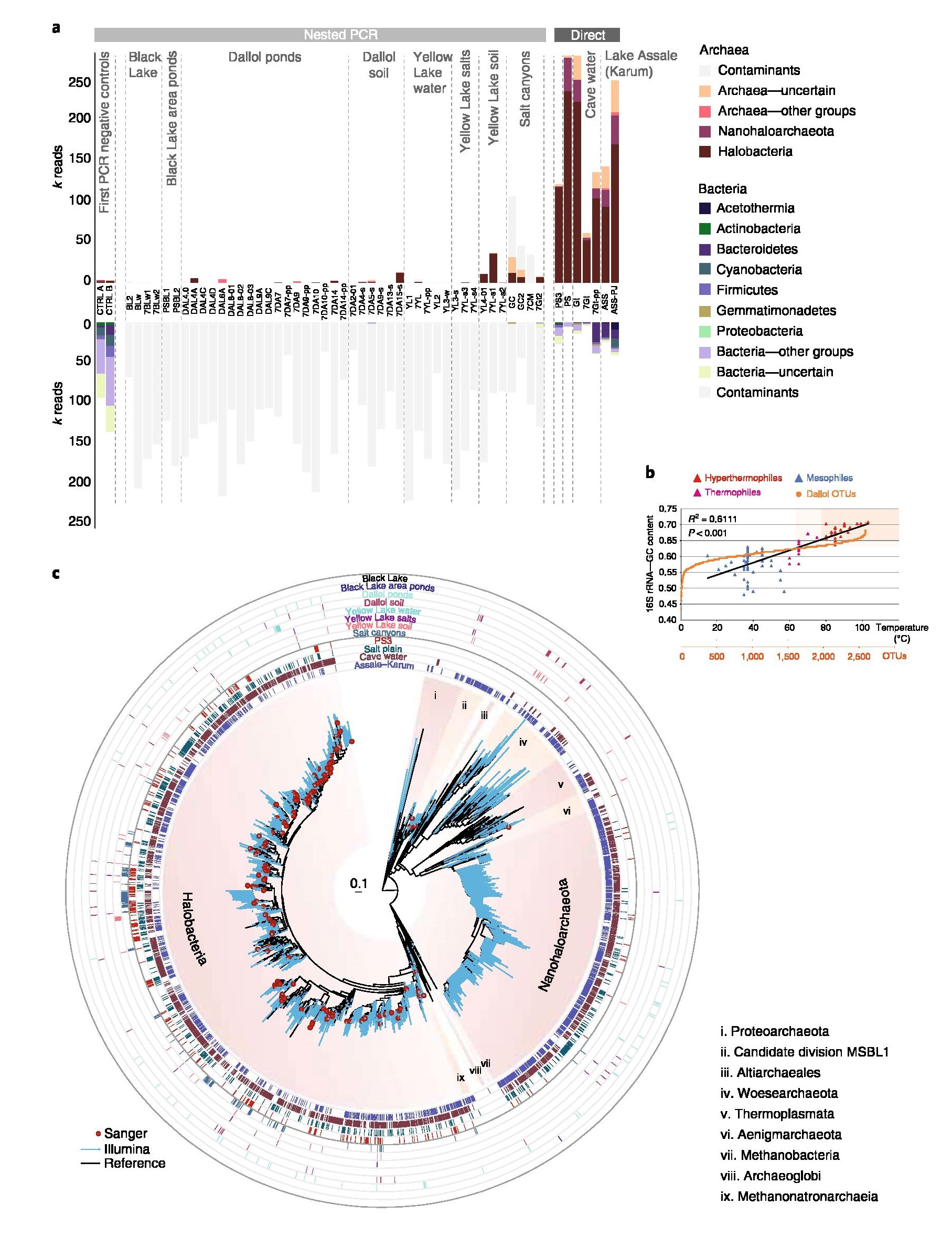
| 18:18, 5 June 2020 | Prokyarotic distribution and diversity at Dallol.jpg (file) |  |
758 KB | Rachelso2020 | The original file was replaced with a higher resolution version so the text shows up more starkly. | 2 |
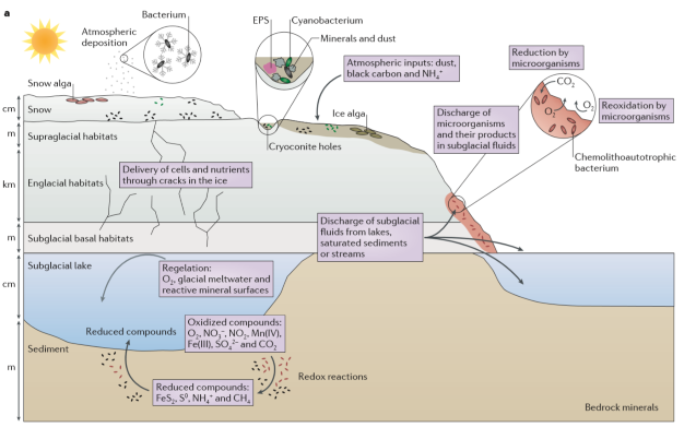
| 20:10, 4 June 2020 | Boeitus et al. 2015 Diagram.png (file) |  |
314 KB | Timothycoston2024 | Modified from: Boetius, A., Anesio, A.M., Deming, J.W., Mikucki, J.A., and Rapp, J.Z., 2015, Microbial ecology of the cryosphere: sea ice and glacial habitats: Nature Reviews, v. 13, p. 677-690. | 1 |
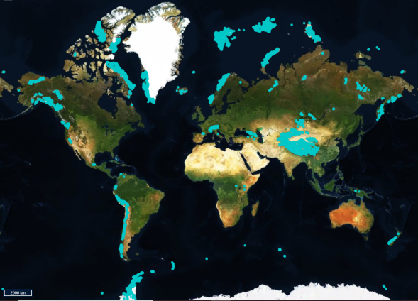
| 20:08, 4 June 2020 | RGI2.png (file) |  |
326 KB | Timothycoston2024 | https://www.glims.org/maps/glims Accessed June 3, 2020 | 1 |
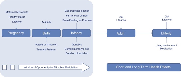
| 09:09, 4 June 2020 | Microbiome acquisition.jpg (file) |  |
56 KB | Nhutran2021 | 1 | |
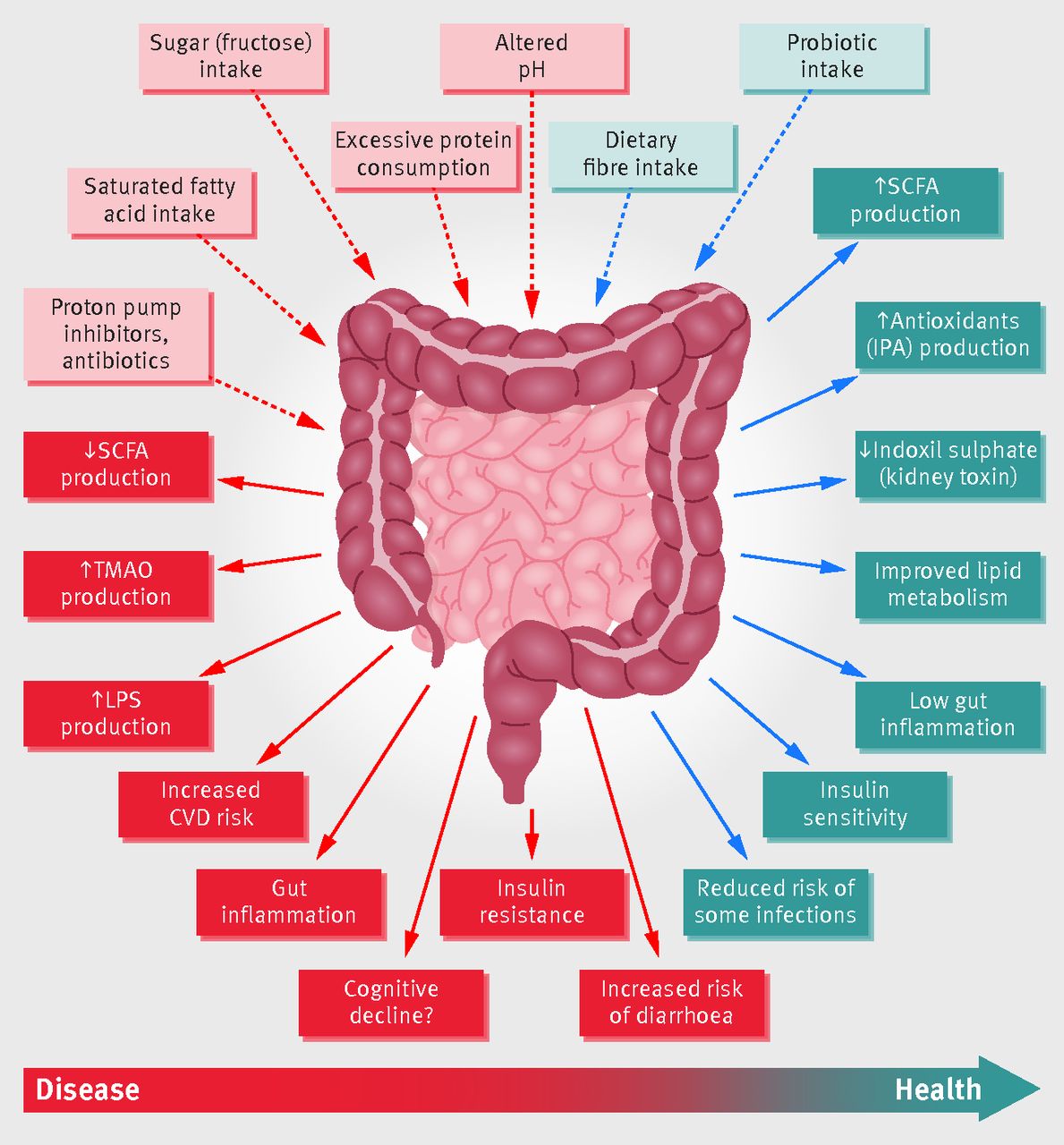
| 08:28, 4 June 2020 | Gut microflora.jpg (file) |  |
180 KB | Nhutran2021 | 1 | |
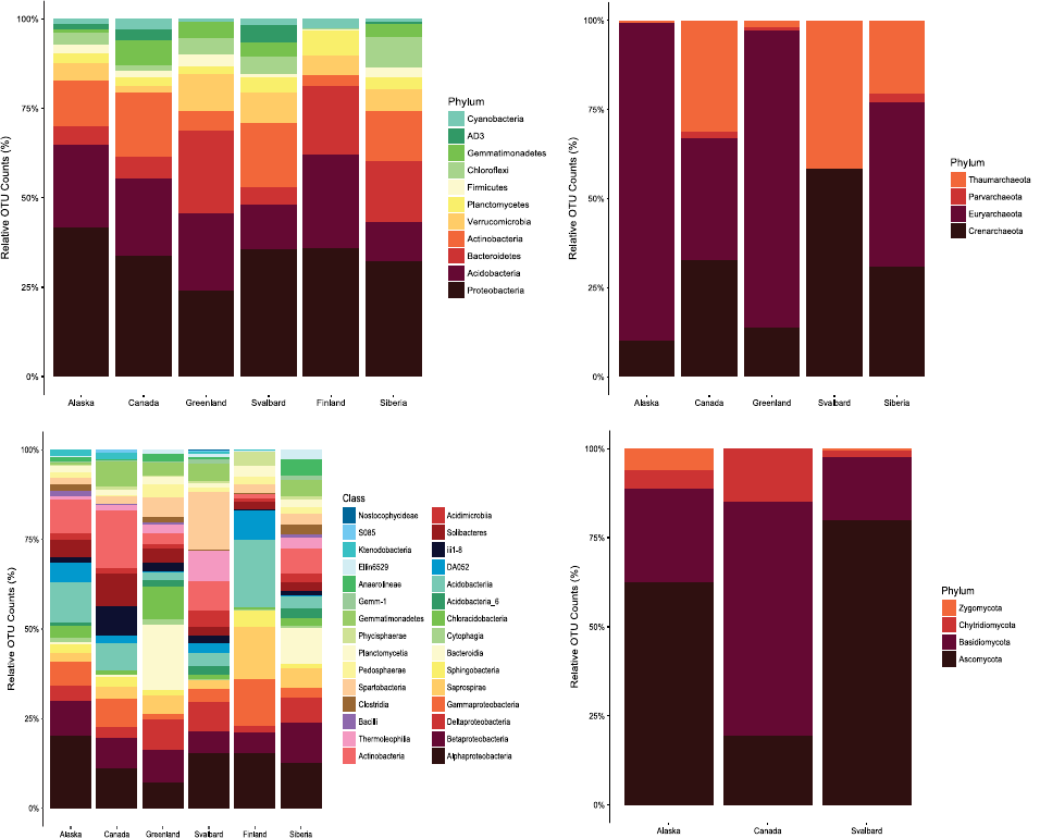
| 22:45, 3 June 2020 | Arcticsoildiversity.png (file) |  |
59 KB | Tiachung-swanson2021 | 1 | |
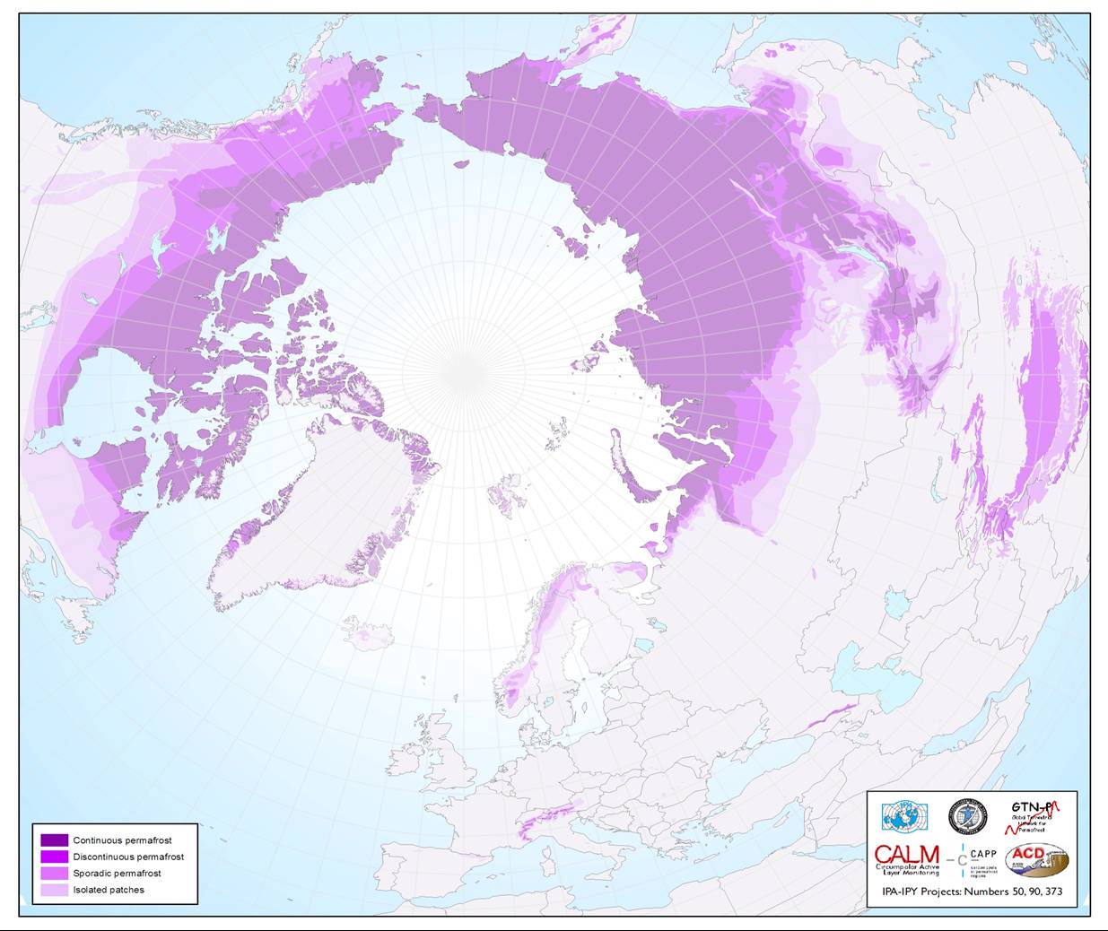
| 17:43, 3 June 2020 | Permafrost map.jpg (file) |  |
132 KB | Tiachung-swanson2021 | 1 | |
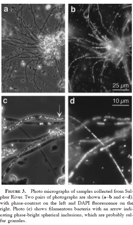
| 17:31, 3 June 2020 | Filamentous cells angert1998.png (file) |  |
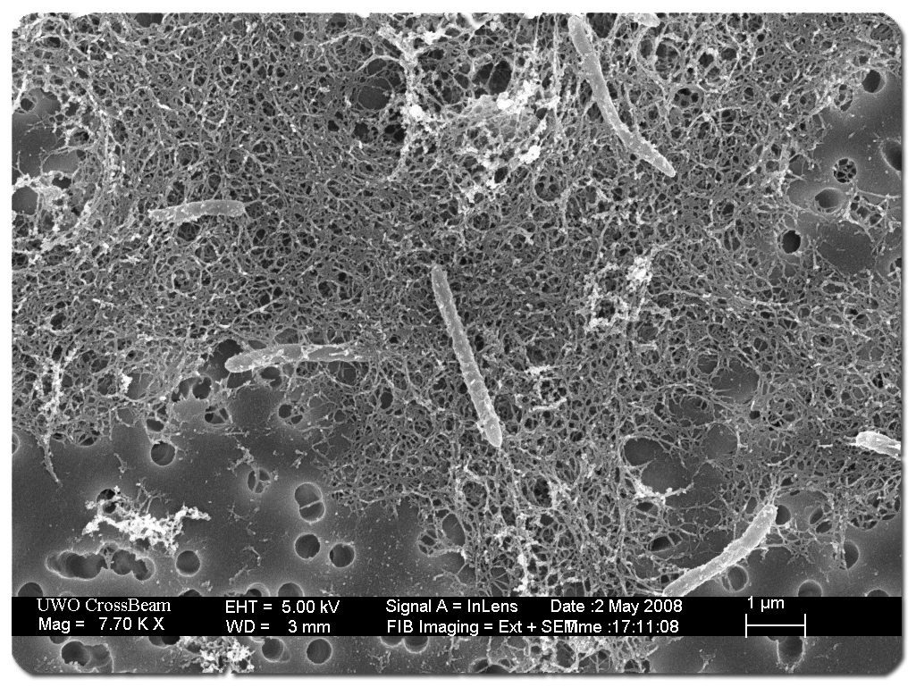
280 KB | Mselensky | Filamentous cells and putative sulfur granules from Sulphur River in Parker Cave, KY, USA. From Angert et al. 1998: https://pubs.geoscienceworld.org/msa/ammin/article-abstract/83/11-12_Part_2/1583/43432 | 1 |
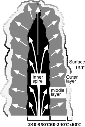
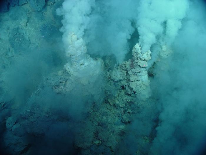
| 14:55, 3 June 2020 | White smoker diagram.gif (file) |  |
23 KB | Carolinewebster2021 | 1 | |
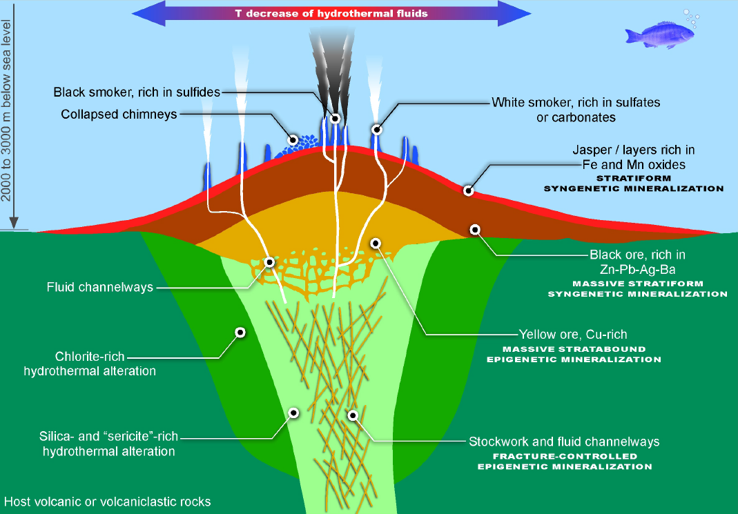
| 14:54, 3 June 2020 | Hydrothermal field diagram.png (file) |  |
380 KB | Carolinewebster2021 | 1 | |
| 14:54, 3 June 2020 | Emi 978 f3.gif (file) |  |
94 KB | Carolinewebster2021 | 1 | |
| 14:53, 3 June 2020 | Emi 978 f2.gif (file) |  |
94 KB | Carolinewebster2021 | 1 | |
| 14:53, 3 June 2020 | Whitesmoker.jpg (file) |  |
36 KB | Carolinewebster2021 | 1 | |
| 10:51, 3 June 2020 | Intro figure 373wiki.jpg (file) |  |
48 KB | Andreagomez-patron2020 | 1 | |
| 09:18, 3 June 2020 | Figure 6 373wiki.jpg (file) |  |
212 KB | Andreagomez-patron2020 | 1 | |
| 09:17, 3 June 2020 | Figure 5 373wiki.png (file) |  |
129 KB | Andreagomez-patron2020 | 1 | |
| 09:17, 3 June 2020 | Figure 4 373wiki.png (file) |  |
91 KB | Andreagomez-patron2020 | 1 | |
| 09:12, 3 June 2020 | Figure 2 373wiki.jpg (file) |  |
325 KB | Andreagomez-patron2020 | 1 | |
| 09:12, 3 June 2020 | Figure 3 373wiki.jpg (file) |  |
185 KB | Andreagomez-patron2020 | 1 | |
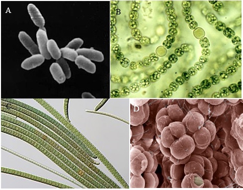
| 09:06, 3 June 2020 | Figure 1 373wiki.png (file) |  |
298 KB | Andreagomez-patron2020 | 1 | |
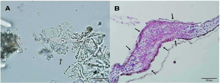
| 01:55, 3 June 2020 | Pseudogymnoascus destructans.png (file) |  |
579 KB | Mselensky | A) Free-living Pseudogymnoascus destructans, the fungal cause of White Nose Syndrome in bats. B) Histopathology slide of P. destructans in bat skin tissue. From Zukal et al. (2016): https://www.nature.com/articles/srep19829 | 1 |
