Streptococcus mutans: More than Oral Pathogens
By: Jordan Wehner
Introduction
History
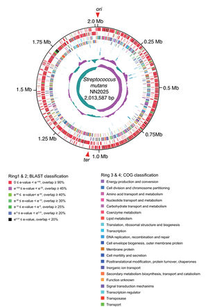
Our genetic code isn’t the only thing that is similar to chimpanzees, we also have oral microbes in common. Specifically, we share the firmicute Streptococcus mutans (Fig. 1). These pathogenic microbes are part of the bacteria kingdom and were first isolated from human cavities (carious lessions) in 1924 by Clarke.XX The microbes received the name S. mutans because Clarke believed that the microbes were a mutant version of streptococcus. These microbes are found in all homosapiens and are the main cause of dental decay. As much as 90 % of the world is affected by this pathogenic microbe. XX
Serotypes
Like many species of bacteria, S. mutans have various strains and serotypes. Serotypes are microorganisms that share a shares a specific substance that cause an immune response.XX Four major serotypes have been discover so far. The serotypes are labeled as c, e, f, and k. The serotype c is considered to be the most common serotype found in plaque, making up about 80 % of the microbes population.XX The other three serotypes (e, f, and k) are less common. Up to 20 % of the S. mutans are considered to be e. The smallest groups, f and k, are thought to make up 5 % of the microbe population. Interestingly, the serotype k has only been isolated from Japanese individuals. Recent research has found that e, f, and k may play a hazardous role in infective endocarditis (e and f) and hemorrhagic strokes (k) even though they only make up about a quarter of the population.XX
Genome Structure
Metabolism
Include some current research in each topic, with at least one figure showing data.
Acid Production and Teeth
Include some current research in each topic, with at least one figure showing data.
S. mutans "Attachment" to Your Heart
Include some current research in each topic, with at least one figure showing data.
The Correlation Between S. mutans and Hemorrhagic Stroke
Overall paper length should be 3,000 words, with at least 3 figures.
Conclusion
Overall paper length should be 3,000 words, with at least 3 figures.
References
Edited by student of Joan Slonczewski for BIOL 238 Microbiology, 2009, Kenyon College.


