The Acquisition, Metabolism, and Pathological Mechanisms Underlying Giardia Lamblia: Difference between revisions
Slevenso3268 (talk | contribs) No edit summary |
Slevenso3268 (talk | contribs) No edit summary |
||
| Line 2: | Line 2: | ||
<br><i>Giardia lamblia</i> is a flagellated protozoan parasite that settles in the small intestine, causing giardiasis. Antonie van Leeuwenhoek, who examined samples of his own diarrheal stool, first documented the [http://en.wikipedia.org/wiki/Apicomplexan_life_cycle trophozoite] form of G. lamblia in 1681. Giardia is confined to the [http://en.wikipedia.org/wiki/Lumen_(anatomy) lumen] of the small intestine, and does not spread through the bloodstream. The main route to infection is ingestion of untreated sewage and contaminated natural waters. It is not only found in humans, but is the most common parasite to infect other animals as well including cats, dogs, birds, cattle, and sheep.[http://www.brianjford.com/Giardia-14-06.pdf/:<sup></sup>] | <br><i>Giardia lamblia</i> is a flagellated protozoan parasite that settles in the small intestine, causing giardiasis. Antonie van Leeuwenhoek, who examined samples of his own diarrheal stool, first documented the [http://en.wikipedia.org/wiki/Apicomplexan_life_cycle trophozoite] form of G. lamblia in 1681. Giardia is confined to the [http://en.wikipedia.org/wiki/Lumen_(anatomy) lumen] of the small intestine, and does not spread through the bloodstream. The main route to infection is ingestion of untreated sewage and contaminated natural waters. It is not only found in humans, but is the most common parasite to infect other animals as well including cats, dogs, birds, cattle, and sheep.[http://www.brianjford.com/Giardia-14-06.pdf/:<sup></sup>] | ||
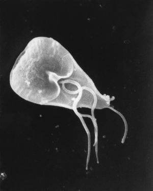
[[Image:GiardiaLamblia.jpg|thumb|300px|right|Electron micrograph of the Giardia lamblia virus|External ultrastructural details displayed by the flagellated protozoan parasite.http://commons.wikimedia.org/wiki/Category:Giardia_lamblia#/media/File:Giardia_lamblia_SEM_8698_lores.jpg/ By Janice Haney Carr at the CDC].[http://commons.wikimedia.org/wiki/Category:Giardia_lamblia#/media/File:Giardia_lamblia_SEM_8698_lores.jpg/<sup>11</sup>]]] | |||
<i>G. lamblia</i> is a flagellated unicellular eukaryotic microorganism that has two major life cycle stages. The cyst that is ingested is inert, which allows it to survive in many different kinds of environmental conditions. However after contact with the acidic environment in the stomach, the cyst will excyst into trophozoites inside of the small intestine. The trophozoites are able to form cysts in the [http://en.wikipedia.org/wiki/Jejunum jejunum] after being exposed to the [http://en.wikipedia.org/wiki/Bile_duct biliary] fluid, which allows transmission of the cyst. | <i>G. lamblia</i> is a flagellated unicellular eukaryotic microorganism that has two major life cycle stages. The cyst that is ingested is inert, which allows it to survive in many different kinds of environmental conditions. However after contact with the acidic environment in the stomach, the cyst will excyst into trophozoites inside of the small intestine. The trophozoites are able to form cysts in the [http://en.wikipedia.org/wiki/Jejunum jejunum] after being exposed to the [http://en.wikipedia.org/wiki/Bile_duct biliary] fluid, which allows transmission of the cyst. | ||
| Line 10: | Line 12: | ||
==Acquisition== | ==Acquisition== | ||
Revision as of 21:59, 11 March 2015
Giardia lamblia is a flagellated protozoan parasite that settles in the small intestine, causing giardiasis. Antonie van Leeuwenhoek, who examined samples of his own diarrheal stool, first documented the trophozoite form of G. lamblia in 1681. Giardia is confined to the lumen of the small intestine, and does not spread through the bloodstream. The main route to infection is ingestion of untreated sewage and contaminated natural waters. It is not only found in humans, but is the most common parasite to infect other animals as well including cats, dogs, birds, cattle, and sheep.

G. lamblia is a flagellated unicellular eukaryotic microorganism that has two major life cycle stages. The cyst that is ingested is inert, which allows it to survive in many different kinds of environmental conditions. However after contact with the acidic environment in the stomach, the cyst will excyst into trophozoites inside of the small intestine. The trophozoites are able to form cysts in the jejunum after being exposed to the biliary fluid, which allows transmission of the cyst.
Researchers have discovered fascinating information about giardia. They have found that giardia can induce apoptosis and disrupt the barrier function in small intestine. G. lamblia also undergoes process that allows the parasite to develop chronic and recurrent infections called antigenic variation, which evades the immune system of the host. G. lamblia also has a very interesting metabolic process. It has little biosynthetic capacity. It lacks mitochondria and its oxidative phosphorylation is not mediated by the cytochrome. As well, it is able to carry out aerobic and anaerobic respiration depending on the amount of oxygen in the environment. Acquisition and metabolism of G. lamblia contribute to the pathological mechanisms of the disease, and how it is able to cause such unpleasant symptoms without actually penetrating the epithelium.
Acquisition
Section 2
All about metabolism
Pathomechanisms
Epithelial Barrier Dysfunction
G. lamblia is able to cause disease without penetrating the epithelium, invading tissues, or entering the bloodstream .
Some patients have with chronic giardiasis have been found to have epithelial barrier dysfunction, which down regulates the tight junction protein claudin 1 and increases epithelial apoptosis. This causes failure of sodium-dependent glucose absorption, which results in active chloride ion secretion. Water then enters the lumen and causes diarrhea. .
In order to see the effect of the infection on epithelial transport and barrier dysfunction, duodenal biopsy specimens from 13 patients with chronic giardiasis and from controls were obtained endoscopically. The resistances of the epithelium and the sub epithelium were found using impedance spectroscopy. As well, researchers performed mucosal morphometry to characterize the tight junction proteins. Overall they found that the patients suffering from giardiasis had a 75% decreased mucosal surface area per unit of serosa area. .
Induced Apoptosis
Giardia lamblia rearranges cytoskeletal proteins that reduces the electrical resistance across the epithelium. To see whether G. lamblia induced enterocyte apoptosis in duodenal epithelilal monolayers, monolayers of duodenal epithelial cells were incubated with G. lamblia trophozoites for a certain amount of time and were treated with or without 120 μM caspase-3 inhibitor (Z-DEVD-FMK). It was found that trophozoite strains NF and S2 induced enterocyte apoptosis within the monolayers, but were inhibited by the Z-DEVD-FMK. It was also found that NF disrupted tight junction ZO-1. Overall, strain-dependent infection of enterocyte apoptosis could contribute to the pathogenesis of G. lamblia. Disrupting tight junction ZO-1 and increasing permeability in a caspase-3 dependent manner could explain the apoptosis effect .
Disruption of the barrier function in the small intestine is T cell independent and increases the permeability of single-layered duodenal epithelium by the phosphoralization of F-actin and of tight junctions in enterocytes .
Further Reading
[Sample link] Ebola Hemorrhagic Fever—Centers for Disease Control and Prevention, Special Pathogens Branch
References
Edited by (your name here), a student of Nora Sullivan in BIOL168L (Microbiology) in The Keck Science Department of the Claremont Colleges Spring 2014.
