The use of antibiotics on Wolbachia as treatment for filarial diseases: Difference between revisions
No edit summary |
No edit summary |
||
| Line 7: | Line 7: | ||
===Localization of ''Wolbachia''=== | ===Localization of ''Wolbachia''=== | ||
''Wolbachia'' is found in stages of larval development in the hypodermal cells of the lateral cords as well as in the embryos of female filarial nematodes. [http://journals.plos.org/plospathogens/article?id=10.1371/journal.ppat.1002351 <sup>2</sup>] | |||
Past studies dating back to the late 1990's and early 2000's have shown that Wolbachia is transmitted by female [http://en.wikipedia.org/wiki/Drosophila_melanogaster ''Drosophila melanogaster''] to offspring by localizing in the posterior female host oocytes, thus increasing its chance to be incorporated into the germline and be passed off to the offspring. More recent studies on ''Drosophila'' have determined the mechanism to be related to the bacteria’s ability to use ''Drosophila'' microtubules, kinesin and dynein in insects to properly segregate to the maternal hosts' posterior germline pole plasma area during oogenesis. [http://journals.plos.org/plospathogens/article?id=10.1371/journal.ppat.0010014 <sup>3</sup>] [http://journals.plos.org/plospathogens/article?id=10.1371/journal.ppat.0030190#s3 <sup>4</sup>] | |||
''Wolbachia'' uses a similar mechanism in both ''Drosophila'' and filarial nematodes. ''Wolbachia'' localizes to maternal centromeres during division. It is also found near the microtubules, which could be interpreted as a possible interaction between ''Wolbachia'' and the microtubules. [http://journals.plos.org/plospathogens/article?id=10.1371/journal.ppat.1002351 <sup>2</sup>] A dynein-based mechanism is used to ''Wolbachia'' make localize to the posterior of the cell. | |||
Removal of ''Wolbachia'' from the filarial nematode host leads to large scale occurrences of apoptosis in the adult germline, and in the somatic cells in certain stages of nematode development leading up to the adult stage.[http://journals.plos.org/plospathogens/article?id=10.1371/journal.ppat.1002351 <sup>5</sup>] The apoptosis in the uterine area results in sterilization of the adult female worms, as the embryos in uterus undergo apoptosis. This considerably reduces the adult worms' ability to spread and cause filarial diseases. Microfilaria are able to remain motile and viable, but are limited in their ability to develop past the L4 larvae stage and thus be able to reach a reproductive age and help the proliferation of more nematodes, an effect caused by apoptosis from the removal of ''Wolbachia'' through antibiotics.[http://journals.plos.org/plospathogens/article?id=10.1371/journal.ppat.1002351 <sup>5</sup>] It was also found that ''Wolbachia'' uses A-P polarity to localize to the posterior of the egg and thus the beginning of the germline. ''Wolbachia'' is needed for the proper A-P polarity in the filarial nematodes, showing the necessity of ''Wolbachia'' to localize in the filarial nematodes' oocytes for normal embryo development. [http://journals.plos.org/plospathogens/article?id=10.1371/journal.ppat.1002351 <sup>2</sup>] Both of these findings could explain how the removal of ''Wolbachia'' leads to sterilization of female filarial nematodes. | |||
===Pathogenesis=== | ===Pathogenesis=== | ||
''Wolbachia'' can create a proinflmmatory response with interaction with specific immunte cells. [http://www.ncbi.nlm.nih.gov/pubmed/23398406 <sup>1</sup>] ''Wolbachia'' creates an ineffective [http://en.wikipedia.org/wiki/Neutrophil_granulocyte neutrophil] response by preventing the disperal of [http://en.wikipedia.org/wiki/Eosinophil_granulocyte eosinophils]. [http://www.ncbi.nlm.nih.gov/pubmed/23398406 <sup>1</sup>]. The 'Wolbachia'' stimulate the production of chemotactic cytokines that determine the neutrophil recruitment. When these neutrophils engulf the ''Wolbachia'' containing filarial nematodes, it stimulates the production of more chemotactic cytokines and thus a greater neutrophil recruitment pull. The large numbers of neutrophils release toxins, resulting in detrimental effects to nearby cells that regulate corneal functions.[http://www.ncbi.nlm.nih.gov/pmc/articles/PMC517527/ <sup>6</sup>] This event shows how ''Wolbachia'' can contribute to onchocerciasis. | |||
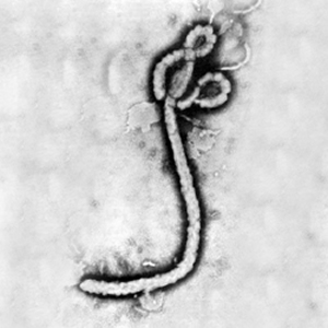
[[Image:Ebola virus 1.jpeg|thumb|300px|right|Electron micrograph of the Ebola Zaire virus. This was the first photo ever taken of the virus, on 10/13/1976. By Dr. F.A. Murphy, now at U.C. Davis, then at the CDC.]] | [[Image:Ebola virus 1.jpeg|thumb|300px|right|Electron micrograph of the Ebola Zaire virus. This was the first photo ever taken of the virus, on 10/13/1976. By Dr. F.A. Murphy, now at U.C. Davis, then at the CDC.]] | ||
<br>At right is a sample image insertion. It works for any image uploaded anywhere to MicrobeWiki. The insertion code consists of: | <br>At right is a sample image insertion. It works for any image uploaded anywhere to MicrobeWiki. The insertion code consists of: | ||
| Line 43: | Line 52: | ||
1. [http://www.ncbi.nlm.nih.gov/pubmed/23398406 Bouchery, T., Lefoulon, E., Karadjian, G., Nieguitsila, A., & Martin, C. (2013). The symbiotic role of Wolbachia in Onchocercidae and its impact on filariasis. Clinical Microbiology & Infection, 19(2), 131–140. Retrieved from 10.1111/1469-0691.12069] | 1. [http://www.ncbi.nlm.nih.gov/pubmed/23398406 Bouchery, T., Lefoulon, E., Karadjian, G., Nieguitsila, A., & Martin, C. (2013). The symbiotic role of Wolbachia in Onchocercidae and its impact on filariasis. Clinical Microbiology & Infection, 19(2), 131–140. Retrieved from 10.1111/1469-0691.12069] | ||
2. [http://journals.plos.org/plospathogens/article?id=10.1371/journal.ppat.1002351 Landmann, F., Voronin, D., Sullivan, W., & Taylor, M. J. (2011). Anti-filarial Activity of Antibiotic Therapy Is Due to Extensive Apoptosis after Wolbachia Depletion from Filarial Nematodes. PLoS Pathog, 7(11), e1002351. Retrieved from http://dx.doi.org/10.1371%2Fjournal.ppat.1002351] | |||
3. (Ferree et al., 2005)[ http://journals.plos.org/plospathogens/article?id=10.1371/journal.ppat.0010014 Ferree, P. M., Frydman, H. M., Li, J. M., Cao, J., Wieschaus, E., & Sullivan, W. (2005). <italic>Wolbachia</italic> Utilizes Host Microtubules and Dynein for Anterior Localization in the <italic>Drosophila</italic> Oocyte. PLoS Pathog, 1(2), e14. Retrieved from http://dx.plos.org/10.1371%2Fjournal.ppat.0010014 | |||
Gillette-Ferguson, I., Hise, A. G., McGarry, H. F., Turner, J., Esposito, A., Sun, Y., … Pearlman, E. (2004). Wolbachia-Induced Neutrophil Activation in a Mouse Model of Ocular Onchocerciasis (River Blindness). Infection and Immunity , 72 (10 ), 5687–5692. doi:10.1128/IAI.72.10.5687-5692.2004 | |||
5. [http://journals.plos.org/plospathogens/article?id=10.1371/journal.ppat.1002351 Landmann, F., Voronin, D., Sullivan, W., & Taylor, M. J. (2011). Anti-filarial Activity of Antibiotic Therapy Is Due to Extensive Apoptosis after Wolbachia Depletion from Filarial Nematodes. PLoS Pathog, 7(11), e1002351. Retrieved from http://dx.doi.org/10.1371%2Fjournal.ppat.1002351] | |||
6. [http://www.ncbi.nlm.nih.gov/pmc/articles/PMC517527/ Gillette-Ferguson, I., Hise, A. G., McGarry, H. F., Turner, J., Esposito, A., Sun, Y., … Pearlman, E. (2004). Wolbachia-Induced Neutrophil Activation in a Mouse Model of Ocular Onchocerciasis (River Blindness). Infection and Immunity , 72 (10 ), 5687–5692. doi:10.1128/IAI.72.10.5687-5692.2004] | |||
[Sample reference] [http://ijs.sgmjournals.org/cgi/reprint/50/2/489 Takai, K., Sugai, A., Itoh, T., and Horikoshi, K. "''Palaeococcus ferrophilus'' gen. nov., sp. nov., a barophilic, hyperthermophilic archaeon from a deep-sea hydrothermal vent chimney". ''International Journal of Systematic and Evolutionary Microbiology''. 2000. Volume 50. p. 489-500.] | [Sample reference] [http://ijs.sgmjournals.org/cgi/reprint/50/2/489 Takai, K., Sugai, A., Itoh, T., and Horikoshi, K. "''Palaeococcus ferrophilus'' gen. nov., sp. nov., a barophilic, hyperthermophilic archaeon from a deep-sea hydrothermal vent chimney". ''International Journal of Systematic and Evolutionary Microbiology''. 2000. Volume 50. p. 489-500.] | ||
Revision as of 07:46, 17 March 2015
Wolbachia is an endosymbiont that lives in many insects and arthropods. It also lives within Brugia malayi, a filarial nematode that can cause lymphatic filariasis, and Onchocerca volvulus, adifferent filarial nematode that causes onchocerciasis. Due to the symbiotic relationship that makes these organisms’ processes so specialized and heavily dependent on each other for survival, treatment of Wolbachia with antibiotics is a possible target for antifilarial activities. Thus recent research looks for new modes of action for novel antibiotics to treat filarial caused diseases to possibly prevent Wolbachia from reaching antibiotic resistance. This field is a rapidly growing research area with many more discoveries to be made and questions to answer.
Symbiotic Relationship
Past research has shown that the removal of Wolbachia from the host filarial nematodes causes antifilarial effects.1 To understand why this event occurs, the evolution of the symbiotic relationship between host and endosymbiont must be understood. This knowledge could lead to new antibiotic treatments as the mechanisms behind the proliferation and transmission of Wolbachia through the life cycle of the host can lead to novel targets for antibiotics.
Localization of Wolbachia
Wolbachia is found in stages of larval development in the hypodermal cells of the lateral cords as well as in the embryos of female filarial nematodes. 2 Past studies dating back to the late 1990's and early 2000's have shown that Wolbachia is transmitted by female Drosophila melanogaster to offspring by localizing in the posterior female host oocytes, thus increasing its chance to be incorporated into the germline and be passed off to the offspring. More recent studies on Drosophila have determined the mechanism to be related to the bacteria’s ability to use Drosophila microtubules, kinesin and dynein in insects to properly segregate to the maternal hosts' posterior germline pole plasma area during oogenesis. 3 4
Wolbachia uses a similar mechanism in both Drosophila and filarial nematodes. Wolbachia localizes to maternal centromeres during division. It is also found near the microtubules, which could be interpreted as a possible interaction between Wolbachia and the microtubules. 2 A dynein-based mechanism is used to Wolbachia make localize to the posterior of the cell. Removal of Wolbachia from the filarial nematode host leads to large scale occurrences of apoptosis in the adult germline, and in the somatic cells in certain stages of nematode development leading up to the adult stage.5 The apoptosis in the uterine area results in sterilization of the adult female worms, as the embryos in uterus undergo apoptosis. This considerably reduces the adult worms' ability to spread and cause filarial diseases. Microfilaria are able to remain motile and viable, but are limited in their ability to develop past the L4 larvae stage and thus be able to reach a reproductive age and help the proliferation of more nematodes, an effect caused by apoptosis from the removal of Wolbachia through antibiotics.5 It was also found that Wolbachia uses A-P polarity to localize to the posterior of the egg and thus the beginning of the germline. Wolbachia is needed for the proper A-P polarity in the filarial nematodes, showing the necessity of Wolbachia to localize in the filarial nematodes' oocytes for normal embryo development. 2 Both of these findings could explain how the removal of Wolbachia leads to sterilization of female filarial nematodes.
Pathogenesis
Wolbachia can create a proinflmmatory response with interaction with specific immunte cells. 1 Wolbachia creates an ineffective neutrophil response by preventing the disperal of eosinophils. 1. The 'Wolbachia stimulate the production of chemotactic cytokines that determine the neutrophil recruitment. When these neutrophils engulf the Wolbachia containing filarial nematodes, it stimulates the production of more chemotactic cytokines and thus a greater neutrophil recruitment pull. The large numbers of neutrophils release toxins, resulting in detrimental effects to nearby cells that regulate corneal functions.6 This event shows how Wolbachia can contribute to onchocerciasis.
At right is a sample image insertion. It works for any image uploaded anywhere to MicrobeWiki. The insertion code consists of:
Double brackets: [[
Filename: Ebola virus 1.jpeg
Thumbnail status: |thumb|
Pixel size: |300px|
Placement on page: |right|
Legend/credit: Electron micrograph of the Ebola Zaire virus. This was the first photo ever taken of the virus, on 10/13/1976. By Dr. F.A. Murphy, now at U.C. Davis, then at the CDC.
Closed double brackets: ]]
Other examples:
Bold
Italic
Subscript: H2O
Superscript: Fe3+
Overall paper length should be 3,000 words, with at least 3 figures with data.
Effectiveness of Antibiotic Treatments
Section 3
Include some current research in each topic, with at least one figure showing data.
Further Reading
[Sample link] Ebola Hemorrhagic Fever—Centers for Disease Control and Prevention, Special Pathogens Branch
References
3. (Ferree et al., 2005)[ http://journals.plos.org/plospathogens/article?id=10.1371/journal.ppat.0010014 Ferree, P. M., Frydman, H. M., Li, J. M., Cao, J., Wieschaus, E., & Sullivan, W. (2005). <italic>Wolbachia</italic> Utilizes Host Microtubules and Dynein for Anterior Localization in the <italic>Drosophila</italic> Oocyte. PLoS Pathog, 1(2), e14. Retrieved from http://dx.plos.org/10.1371%2Fjournal.ppat.0010014 Gillette-Ferguson, I., Hise, A. G., McGarry, H. F., Turner, J., Esposito, A., Sun, Y., … Pearlman, E. (2004). Wolbachia-Induced Neutrophil Activation in a Mouse Model of Ocular Onchocerciasis (River Blindness). Infection and Immunity , 72 (10 ), 5687–5692. doi:10.1128/IAI.72.10.5687-5692.2004
Edited by Nitin Kuppanda, a student of Suzanne Kern in BIOL168L (Microbiology) in The Keck Science Department of the Claremont Colleges Spring 2015.

