Transmission of T. Forsythia to humans from a dog bite: Difference between revisions
Leenhoutsm (talk | contribs) |
Leenhoutsm (talk | contribs) |
||
| Line 26: | Line 26: | ||
==Abscesses== | ==Abscesses== | ||
Normally found in oral cavities, bacteria such as T. forsythia have been cultured in other locations on the human body. | Normally found in oral cavities, bacteria such as T. forsythia have been cultured in other locations on the human body. These bacteria are found in abscesses that form from a cut or puncture wound caused by an infected tooth. An instance where this could possibly occur would be if a dog bites a human and has a periodontal disease. The dog is most likely playing host to T. Forsythia and other red complex bacteria. These microbes are hiding in the plaque biofilm that has built up along the periodontal tissues. When an infected dog bites a human, the canine’s teeth puncture the epidermis of that person, and pass along the bacteria to a new host. From there, T. Forsythia infects host cell epithelium and the underlying sebaceous glands. Members of the red complex are detected in and are the cause of what are known as acute periradicular abscesses. | ||
Abscesses are an accumulation of exudate (fluid emitted through a wound) consisting of bacteria, bacterial by-products, inflammatory cells, lysed inflammatory cells, and the content of those cells (Ozebek, et. al, 2010). When these bacteria enter the surrounding epithelial cells, the immune system is unable to suppress the invasion. Abscesses are caused be breaks or punctures in the skin. Bacteria are able to get under the skin and infect the sebaceous and sweat glands underneath the epidermis. This causes an inflammatory response as the body attempts to rid itself of the pathogens. The inflammatory response causes a number of symptoms that are key in properly diagnosing abscesses. In the middle of an abscess, there is liquefaction and an aggregate of dead cells. Along with the liquid and dead cells are bacteria and other debris that are difficult to remove. As time goes on the infected area begins to grow, which creates tension under the skin and greater inflammation characterized by redness, swelling, warmth of the area, and pain to the touch. Pressure from this tension causes the pain. Meanwhile, the bacteria are infecting the sebaceous glands and helping to create pus. This pus is a buildup of dead white blood cells that were attempting to stop the spread of infection along with other dead epithelial cells and debris. | |||
In abscesses, knowing the specific microbe that has caused the infection is important to treating it. Knowing what the microbe is and where it came from is key in prescribing the proper antibiotic. T. forsythia has been known to be most susceptible to the following antibiotics: amoxicillin, clavulanate, ampicillin, doxycycline, tetracycline, and clindamycin (Tanner & Izard, 2006), with amoxicillin and clavulanate being the most effective. Abscesses are often mistaken for cellulitis, so this is another reason that proper detection and identification of the correct bacterium is so crucial. Often with an abscess infection; antibiotics are not enough to completely cure these infections. They tend to need to be incised by a medical professional so they can drain, because drainage does not occur naturally. In extreme cases, abscesses will need to be removed surgically if they have damaged the surrounding tissues badly enough. It is necessary to have abscess treated as soon as possible because there is the possibility that they can begin to infect deeper tissues and possibly enter the bloodstream. Patients with compromised immune systems are especially at risk due to the fact that they do not have the proper defense systems to ward off infections. | |||
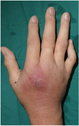
[[Image:1375880716.png|thumb|300px|right|Image of a severely infected abscess. Swelling has begun, along with redness surrounding the injury. [http://www.handsurgery.sg/hand-infection.html].]] | [[Image:1375880716.png|thumb|300px|right|Image of a severely infected abscess. Swelling has begun, along with redness surrounding the injury. [http://www.handsurgery.sg/hand-infection.html].]] | ||
Revision as of 03:58, 24 April 2016
Introduction
T. forsythia is an infectious microbe that is found in the oral cavities of mammals. It is an infectious agent associated with the periodontal diseases of gingivitis and periodontal disease. Gingivitis is characterized by tissue inflammation, and periodontitis is characterized by alveolar bone resorption and bone loss. These diseases/infections become present when plaque buildup is great and allow T. forsythia to have a place to live and reproduce. In the case of dogs, it is common for them to have problems with periodontal diseases due to the fact that they cannot brush their teeth. This poses a problem to humans that are bitten by canines with plaque buildup because they run the risk of being exposed to this microbe. []
Tannerella forsythia

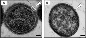
T. Forsythia is a bacterium found within the mouths of mammals and primates. It is mostly found in the oral cavities of humans, dogs, cats, and monkeys. When a gram stain is applied, it is known to be Gram-negative and survives under anaerobic conditions. T. Forsythia only appears in culture from the mouths of mammals with periodontal diseases. These diseases include gingivitis, which is characterized by tissue inflammation, and periodontitis, which is characterized by alveolar bone resorption and bone loss. This bacterium is labeled under the species Bacteroidetes, which led to its original name, Bacteroides forsythia. It has a short, rod-shaped body, and its lack of a flagellum or pili make it non-motile. This lack of mobility or any sort of attachment appendage causes it to be reliant on a biofilm in the form of plaque for it to attach to the surface of teeth or other periodontal tissues. They also have an S-layer comprised of glycoprotein that protects both the inner and outer membranes of the cell. []
Red Complex
T. Forsythia is a part of a group of microbes known as the “Red Complex.” This group of bacteria consists of T. Forsythia, Treponema denticola, and Porphyromonas gingivalis. All of these bacteria are typically found in the oral cavities of mammals. They all are the cause of periodontal diseases such as periodontitis and gingivitis. One of the symptoms and initial clues that these bacteria are present in the periodontal tissues is swelling, inflammation, and bleeding when probed this is where the name Red Complex comes from. Red complex bacteria grow and live as surface attached biofilm plaque in the subgingival area surrounding and underneath the teeth (Homma). T. forsythia, other red complex bacteria and their byproducts are recognized by the host’s immune system and leads to the secretion of cytokines that recruit phagocytic cells such as neutrophils and macrophages. Recognition of any of these bacteria causes a release of antimicrobial peptides for bacterial killing (Homma). In response, the body produces uncontrolled inflammation that results in tissue destruction and increased periodontal pocket depth (Homma). The red, inflamed tissue that results is how the red complex gets its namesake. There is also the possibility for the destruction of tooth-supporting tissue and bone/tooth loss. If any of these symptoms are present in your dog, it is recommended that you take them to them a veterinarian because any members of the Red Complex are easily transmittable from organism to organism. []
Culture
T. Forsythia is a difficult microbe to culture because it is anaerobic. Since they require environments without Oxygen, these cultures are difficult and expensive to create. It is relatively slow growing and requires fastidious growth conditions. When cultured from bite wounds inflicted by dogs, T. Forsythia was shown to grow best in N-Acetylmuramic acid deficient conditions. N-Acetlymuramic acid is an ether of lactic acid and N-Acetylglucosamine, and is often found in the peptidoglycan of bacterial cell walls. However, when T. Forsythia was cultured from human mouths, it was found that they did not grow under conditions with N-Acetylmuramic acid present. In culture, the colonies are very small and are opaque colored. T. Forsythia has also been know to be able to metabolize a range of substrates and like other enteric Bacteroides species, hydrolyzes aesculin. []
Periodontal Diseases in Canines

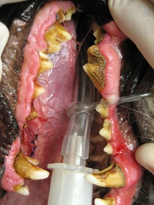
These infections are some of the most commons in dogs as well as cats today. They first start to form when food buildup occurs on a dog’s teeth, and is not properly wiped away. This buildup then transforms into plaque and accumulates along the dog’s gum line. Plaque can then transform into calculus when combined with saliva and other minerals that are common in a dog’s mouth. Calculus is the hardening of plaque, and because this occurs along the gum line, causes gun irritation and inflammation. Theses buildups are breeding grounds for bacteria that live in oral cavities. Microbes from the Bacteroidetes, Actinomyces, and the Streptococcus families all enjoy these kinds of conditions and have been found in cultures from canine tooth plaque. These symptoms are triggers for gingivitis and periodontal disease. In extreme cases, the calculus will build up under the gum and cause separation between the gum and the tooth. This leads to bone degeneration, tooth loss, and pus formation, and the canine will officially have an irreversible periodontal disease. In diagnosing periodontal diseases, a veterinarian will probe the infected area of the dog’s mouth, and if more than two millimeters of space are found between the tooth and gum of the affected area, then the canine officially has a periodontal disease. Certain canine breeds are more likely to be affected then others. The toy breeds are born with crowded teeth, which make it more likely that they will struggle with plaque buildup. Dogs that groom themselves also run a greater chance of getting an infection due to the nature of some areas of the body they have to clean. Poor nutrition can also be a problem because the dog may not be receiving enough nutrients to ward off disease, or they may be receiving those that are helping plaque to harden. The best way to prevent your dog from receiving a periodontal infection is to brush their teeth regularly and wipe their mouths after they eat to remove any food buildup. (PetMD) []
S-layer and BspA
Like all bacteria, T. forsythia has an outer layer known as a S-layer. This S-layer is different from other prokaryotes though. Biochemical and molecular genetics studies have shown that the structure of a T-forsythia S-layer is unique (Yoo et al., 2007). According to Lee et al. (2006), it consists of two large separate glycoproteins that exhibit little to no homology to other S-layer proteins or any other glycoproteins from other prokaryotic organisms. Interestingly, Sabet et al. proved that the S-layer of T. Forsythia is a putative virulence factor mediating certain cellular interactions such as hemagglutination, adherence, invasion, and protective immunity. It is also believed that S-layer proteins play a role in attachment to the tissues and membranes of the oral cavities of infected individuals.
Another known virulence factor on the surface of a T. forsythia cell is BspA (Yoo et al., 2007). BspA is an outer cell surface-associated protein that has been shown to have a role in the pathogenesis of T. forsythia. Honma et al. (2001) proved that BspA is involved with the binding to fibronectin and other extracellular matrix proteins of a host epithelial cell. “It was also observed that BspA mediates epithelial cell attachment and invasion (Inagaki et al., 2006), alveolar bone loss in mice (Sharma et al., 2005a), and coaggregation with Fusobacterium nucleatum (Sharma et al., 2005b). F. nucleatum is an important microbe in dental plaque biofilm formation. Oral cavity bacteria do not have the necessary attachment machinery to latch onto a oral tissue epithelium on their own, so they rely on proteins such as those in the S-layer and BspA to help with attachment as well as biofilms to help keep them in place.
Other studies have shown that protein phosphorylation and cytoskeletal remodeling are necessary for invasion of host epithelial cells (Inagaki et al., 2006). This allows the cell to evade initial detection from the host’s immune system. Terminal sugars known as Sialic acids are commonly present on the external surface of glycoproteins as well as the cell surface of T. forsythia (Angata & Varki 2002; Varki 1997). Sialic acid assists in a variety of biological functions such as cell-cell interactions, stabilizing glycoprotein structure and masking ligand-binding receptors Angata & Varki, 2002; Varki, 1997; Bradshaw et al., 1998). These sialic acids are important to biofilm formation and adhesion to periodontal tissues. The enzyme, NanH sialidase, synthesizes sialic acid and is an important molecular structure when trying to develop a targeted antibiotic for T. forsythia. By turning on the salidase inhibitor, there is the potential to prevent biofilms from forming on the surface of periodontal tissues. With this ability comes the possibility of limiting periodontal diseases from occurring and prevents transmission from canine to humans.
Abscesses
Normally found in oral cavities, bacteria such as T. forsythia have been cultured in other locations on the human body. These bacteria are found in abscesses that form from a cut or puncture wound caused by an infected tooth. An instance where this could possibly occur would be if a dog bites a human and has a periodontal disease. The dog is most likely playing host to T. Forsythia and other red complex bacteria. These microbes are hiding in the plaque biofilm that has built up along the periodontal tissues. When an infected dog bites a human, the canine’s teeth puncture the epidermis of that person, and pass along the bacteria to a new host. From there, T. Forsythia infects host cell epithelium and the underlying sebaceous glands. Members of the red complex are detected in and are the cause of what are known as acute periradicular abscesses.
Abscesses are an accumulation of exudate (fluid emitted through a wound) consisting of bacteria, bacterial by-products, inflammatory cells, lysed inflammatory cells, and the content of those cells (Ozebek, et. al, 2010). When these bacteria enter the surrounding epithelial cells, the immune system is unable to suppress the invasion. Abscesses are caused be breaks or punctures in the skin. Bacteria are able to get under the skin and infect the sebaceous and sweat glands underneath the epidermis. This causes an inflammatory response as the body attempts to rid itself of the pathogens. The inflammatory response causes a number of symptoms that are key in properly diagnosing abscesses. In the middle of an abscess, there is liquefaction and an aggregate of dead cells. Along with the liquid and dead cells are bacteria and other debris that are difficult to remove. As time goes on the infected area begins to grow, which creates tension under the skin and greater inflammation characterized by redness, swelling, warmth of the area, and pain to the touch. Pressure from this tension causes the pain. Meanwhile, the bacteria are infecting the sebaceous glands and helping to create pus. This pus is a buildup of dead white blood cells that were attempting to stop the spread of infection along with other dead epithelial cells and debris.
In abscesses, knowing the specific microbe that has caused the infection is important to treating it. Knowing what the microbe is and where it came from is key in prescribing the proper antibiotic. T. forsythia has been known to be most susceptible to the following antibiotics: amoxicillin, clavulanate, ampicillin, doxycycline, tetracycline, and clindamycin (Tanner & Izard, 2006), with amoxicillin and clavulanate being the most effective. Abscesses are often mistaken for cellulitis, so this is another reason that proper detection and identification of the correct bacterium is so crucial. Often with an abscess infection; antibiotics are not enough to completely cure these infections. They tend to need to be incised by a medical professional so they can drain, because drainage does not occur naturally. In extreme cases, abscesses will need to be removed surgically if they have damaged the surrounding tissues badly enough. It is necessary to have abscess treated as soon as possible because there is the possibility that they can begin to infect deeper tissues and possibly enter the bloodstream. Patients with compromised immune systems are especially at risk due to the fact that they do not have the proper defense systems to ward off infections.

References
"PetMD." Dog Gum Disease. PetMD, n.d. Web. 21 Apr. 2016.
[Lee SW, Sabet M, Um HS, Yang J, Kim HC & Zhu W (2006) Identification and characterization of the genes encoding a unique surface (S-) layer of Tannerella forsythia. Gene 371:102–111.]
[Sharma A, Inagaki S, Honma K, Sfintescu C, Baker PJ & Evans RT (2005a) Tannerella forsythia-induced alveolar bone loss in mice involves leucine-rich-repeat BspA protein. J Dent Res 84:462–467.]
[Angata, T. & Varki, A. (2002). Chemical diversity in the sialic acids and related a-keto acids: an evolutionary perspective. Chem Rev 102, 439–470.]
[Varki, A. (1997). Sialic acids as ligands in recognition phenomena. FASEB J 11, 248–255.]
[Inagaki, S., Onishi, S., Kuramitsu, H. K. & Sharma, A. (2006). Porphyromonas gingivalis vesicles enhance attachment, and the leucine-rich repeat BspA protein is required for invasion of epithelial cells by ‘‘Tannerella forsythia’’. Infect Immun 74, 5023–5028.]
[Sharma A, Inagaki S, Sigurdson W & Kuramitsu HK (2005b) Synergy between Tannerella forsythia and Fusobacterium nucleatum in biofilm formation. Oral Microbiol Immunol 20:39–42. Sharma A, S]
At right is a sample image insertion. It works for any image uploaded anywhere to MicrobeWiki.
The insertion code consists of:
Double brackets: [[
Filename: PHIL_1181_lores.jpg
Thumbnail status: |thumb|
Pixel size: |300px|
Placement on page: |right|
Legend/credit: Electron micrograph of the Ebola Zaire virus. This was the first photo ever taken of the virus, on 10/13/1976. By Dr. F.A. Murphy, now at U.C. Davis, then at the CDC.
Closed double brackets: ]]
Other examples:
Bold
Italic
Subscript: H2O
Superscript: Fe3+
Introduce the topic of your paper. What is your research question? What experiments have addressed your question? Applications for medicine and/or environment?
Sample citations: [1]
[2]
A citation code consists of a hyperlinked reference within "ref" begin and end codes.
