Transmission of T. Forsythia to humans from a dog bite: Difference between revisions
Leenhoutsm (talk | contribs) |
Leenhoutsm (talk | contribs) |
||
| Line 15: | Line 15: | ||
==References== | ==References== | ||
[http://www.kingwestvets.com/brush-dogs-teeth/ Jullian, Tim. "How to Brush Dogs' Teeth | Step by Step Guide | King West Vets." King West Vets. King West Vets Veterinary Clinic, 15 Jan. 2013. Web. 21 Apr. 2016.] | [http://www.kingwestvets.com/brush-dogs-teeth/ Jullian, Tim. "How to Brush Dogs' Teeth | Step by Step Guide | King West Vets." King West Vets. King West Vets Veterinary Clinic, 15 Jan. 2013. Web. 21 Apr. 2016.] | ||
[http://www.petmd.com/dog/conditions/mouth/c_multi_periodontal_disease?page=show "PetMD." Dog Gum Disease. PetMD, n.d. Web. 21 Apr. 2016.] | [http://www.petmd.com/dog/conditions/mouth/c_multi_periodontal_disease?page=show "PetMD." Dog Gum Disease. PetMD, n.d. Web. 21 Apr. 2016.] | ||
<br>At right is a sample image insertion. It works for any image uploaded anywhere to MicrobeWiki.<br><br>The insertion code consists of: | <br>At right is a sample image insertion. It works for any image uploaded anywhere to MicrobeWiki.<br><br>The insertion code consists of: | ||
<br><b>Double brackets:</b> [[ | <br><b>Double brackets:</b> [[ | ||
Revision as of 23:51, 21 April 2016
Tannerella forsythia

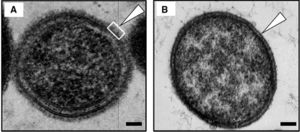
T. Forsythia is a bacterium found within the mouths of mammals and primates. It is mostly found in the oral cavities of humans, dogs, cats, and monkeys. When a gram stain is applied, it is known to be Gram-negative and survives under anaerobic conditions. T. Forsythia only appears in culture from the mouths of mammals with periodontal diseases. These diseases include gingivitis, which is characterized by tissue inflammation, and periodontitis, which is characterized by alveolar bone resorption and bone loss. This bacterium is labeled under the species Bacteroidetes, which led to its original name, Bacteroides forsythia. It has a short, rod-shaped body, and its lack of a flagellum or pili make it non-motile. This lack of mobility or any sort of attachment appendage causes it to be reliant on a biofilm in the form of plaque for it to attach to the surface of teeth or other periodontal tissues. They also have an S-layer comprised of glycoprotein that protects both the inner and outer membranes of the cell. []
Red Complex
T. Forsythia is a part of a group of microbes known as the “Red Complex.” This group of bacteria consists of T. Forsythia, Treponema denticola, and Porphyromonas gingivalis. All of these bacteria are typically found in the oral cavities of mammals. They all are the cause of periodontal diseases such as periodontitis and gingivitis. One of the symptoms and initial clues that these bacteria are present in the periodontal tissues is swelling, inflammation, and bleeding when probed this is where the name Red Complex comes from. If any of these symptoms are present in your dog, it is recommended that you take them to them a veterinarian because any members of the Red Complex are easily transmittable from organism to organism. []
Culture
T. Forsythia is a difficult microbe to culture because it is anaerobic. Since they require environments without Oxygen, these cultures are difficult and expensive to create. It is relatively slow growing and requires fastidious growth conditions. When cultured from bite wounds inflicted by dogs, T. Forsythia was shown to grow best in N-Acetylmuramic acid deficient conditions. N-Acetlymuramic acid is an ether of lactic acid and N-Acetylglucosamine, and is often found in the peptidoglycan of bacterial cell walls. However, when T. Forsythia was cultured from human mouths, it was found that they did not grow under conditions with N-Acetylmuramic acid present. In culture, the colonies are very small and are opaque colored. T. Forsythia has also been know to be able to metabolize a range of substrates and like other enteric Bacteroides species, hydrolyzes aesculin. []
Periodontal Diseases in Canines

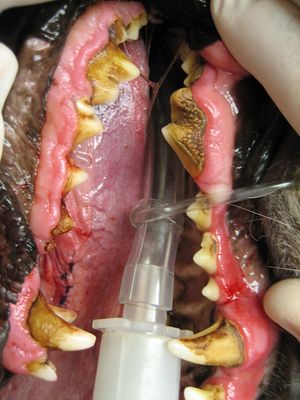
These infections are some of the most commons in dogs as well as cats today. They first start to form when food buildup occurs on a dog’s teeth, and is not properly wiped away. This buildup then transforms into plaque and accumulates along the dog’s gum line. Plaque can then transform into calculus when combined with saliva and other minerals that are common in a dog’s mouth. Calculus is the hardening of plaque, and because this occurs along the gum line, causes gun irritation and inflammation. These symptoms are triggers for gingivitis and periodontal disease. In extreme cases, the calculus will build up under the gum and cause separation between the gum and the tooth. This leads to bone degeneration, tooth loss, and pus formation, and the canine will officially have an irreversible periodontal disease. In diagnosing periodontal diseases, a veterinarian will probe the infected area of the dog’s mouth, and if more than two millimeters of space are found between the tooth and gum of the affected area, then the canine officially has a periodontal disease. Certain canine breeds are more likely to be affected then others. The toy breeds are born with crowded teeth, which make it more likely that they will struggle with plaque buildup. Dogs that groom themselves also run a greater chance of getting an infection due to the nature of some areas of the body they have to clean. Poor nutrition can also be a problem because the dog may not be receiving enough nutrients to ward off disease, or they may be receiving those that are helping plaque to harden. []
References
"PetMD." Dog Gum Disease. PetMD, n.d. Web. 21 Apr. 2016.
At right is a sample image insertion. It works for any image uploaded anywhere to MicrobeWiki.
The insertion code consists of:
Double brackets: [[
Filename: PHIL_1181_lores.jpg
Thumbnail status: |thumb|
Pixel size: |300px|
Placement on page: |right|
Legend/credit: Electron micrograph of the Ebola Zaire virus. This was the first photo ever taken of the virus, on 10/13/1976. By Dr. F.A. Murphy, now at U.C. Davis, then at the CDC.
Closed double brackets: ]]
Other examples:
Bold
Italic
Subscript: H2O
Superscript: Fe3+
Introduce the topic of your paper. What is your research question? What experiments have addressed your question? Applications for medicine and/or environment?
Sample citations: [1]
[2]
A citation code consists of a hyperlinked reference within "ref" begin and end codes.
