User talk:CrowJ: Difference between revisions
(The History and Evolution of the Zika Virus) |
No edit summary |
||
| Line 1: | Line 1: | ||
<!-- Do not edit this line-->{{Curated}} | <!-- Do not edit this line-->{{Curated}} | ||
==Section== | ==Section== | ||
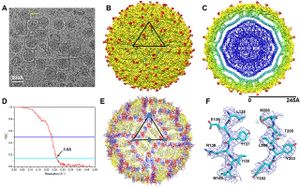
[[Image: | [[Image:Zika fig1.jpg|thumb|300px|right|Cryo-electron micrograph of the Zika virus. This was the first resolution of the virus published in <i>Science</i> on March 31<sup>st</sup>, 2016 by Sirohi et al. The figure shows a representation of virion phenotypes (A); "A surface-shaded depth cued representation of ZIKV viewed down the icosahedral two-fold axis" (B); A cross section of the virus (C) A plot of the Fourier shell coefficient (D); A representation of the protein backbone of the virus (E); A visualization of several amino acids in a backbone protein (F)[http://science.sciencemag.org/content/early/2016/03/30/science.aaf5316].]] | ||
< | |||
< | |||
<br>Introduce the topic of your paper. What is your research question? What experiments have addressed your question? Applications for medicine and/or environment?<br> | |||
<br> Zika virus (ZIKV) is a member of the virus family Flaviviridae and the genus Flavivirus. It is spread by Aedes mosquitoes, for instance A. aegypti. The virus was first isolated in the Zika Forest of Uganda, in 1947. Zika virus is related to other Flaviviruses: dengue, yellow fever, Japanese encephalitis, and West Nile viruses. The virus causes a fever, often with none or only mild symptoms, and go unrecognized in the infected host. In 2013, the virus spread eastward across the Pacific Ocean to French Polynesia, New Caledonia, the Cook Islands, and Easter Island, and in 2015 to Mexico, Central America, the Caribbean, and South America, notably in Brazil. As of 2016, the illness cannot be prevented by medications or vaccines. Zika is thought to spread from an infected pregnant women to the baby. The CDC has linked infant infection with microcephaly and other severe brain problems. Zika infections in adults has been shown to cause Guillain-Barré syndrome. The purpose of this paper is to examine the evolution, the development of virulence in the virus and the link between severe autoimmune-neurological syndrome in infected human hosts and the virus. | |||
Introduce the topic of your paper. What is your research question? What experiments have addressed your question? Applications for medicine and/or environment?<br> | |||
Sample citations: <ref>[http://www.plosbiology.org/article/fetchObject.action?uri=info%3Adoi%2F10.1371%2Fjournal.pbio.1000005&representation=PDF Hodgkin, J. and Partridge, F.A. "<i>Caenorhabditis elegans</i> meets microsporidia: the nematode killers from Paris." 2008. PLoS Biology 6:2634-2637.]</ref> | Sample citations: <ref>[http://www.plosbiology.org/article/fetchObject.action?uri=info%3Adoi%2F10.1371%2Fjournal.pbio.1000005&representation=PDF Hodgkin, J. and Partridge, F.A. "<i>Caenorhabditis elegans</i> meets microsporidia: the nematode killers from Paris." 2008. PLoS Biology 6:2634-2637.]</ref> | ||
<ref>[http://www.ncbi.nlm.nih.gov/pmc/articles/PMC3847443/ Bartlett et al.: Oncolytic viruses as therapeutic cancer vaccines. Molecular Cancer 2013 12:103.]</ref> | <ref>[http://www.ncbi.nlm.nih.gov/pmc/articles/PMC3847443/ Bartlett et al.: Oncolytic viruses as therapeutic cancer vaccines. Molecular Cancer 2013 12:103.]</ref> | ||
Revision as of 20:35, 21 April 2016
Section

Zika virus (ZIKV) is a member of the virus family Flaviviridae and the genus Flavivirus. It is spread by Aedes mosquitoes, for instance A. aegypti. The virus was first isolated in the Zika Forest of Uganda, in 1947. Zika virus is related to other Flaviviruses: dengue, yellow fever, Japanese encephalitis, and West Nile viruses. The virus causes a fever, often with none or only mild symptoms, and go unrecognized in the infected host. In 2013, the virus spread eastward across the Pacific Ocean to French Polynesia, New Caledonia, the Cook Islands, and Easter Island, and in 2015 to Mexico, Central America, the Caribbean, and South America, notably in Brazil. As of 2016, the illness cannot be prevented by medications or vaccines. Zika is thought to spread from an infected pregnant women to the baby. The CDC has linked infant infection with microcephaly and other severe brain problems. Zika infections in adults has been shown to cause Guillain-Barré syndrome. The purpose of this paper is to examine the evolution, the development of virulence in the virus and the link between severe autoimmune-neurological syndrome in infected human hosts and the virus.
Introduce the topic of your paper. What is your research question? What experiments have addressed your question? Applications for medicine and/or environment?
Sample citations: [1]
[2]
A citation code consists of a hyperlinked reference within "ref" begin and end codes.
Section 1
Include some current research, with at least one figure showing data.
Every point of information REQUIRES CITATION using the citation tool shown above.
Section 2
Include some current research, with at least one figure showing data.
Section 3
Include some current research, with at least one figure showing data.
Section 4
Conclusion
References
Authored for BIOL 238 Microbiology, taught by Joan Slonczewski, 2016, Kenyon College.
