West Nile Virus and Acute Flaccid Paralysis: Difference between revisions
From MicrobeWiki, the student-edited microbiology resource
| Line 2: | Line 2: | ||
<br>By Kate Lang<br> | <br>By Kate Lang<br> | ||
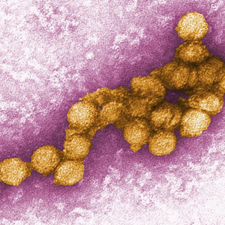
[[Image:flaviviridae1.jpeg|thumb|300px|right|Electron micrograph image of West Nile virus. This image is made public courteous of the Centers for Disease Control and Prevention, specifically the Viral Special Pathogens Branch.]] | [[Image:flaviviridae1.jpeg|thumb|300px|right|Electron micrograph image of West Nile virus. This image is made public courteous of the Centers for Disease Control and Prevention, specifically the Viral Special Pathogens Branch.]] | ||
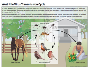
[[Image:WNVtransmissioncycle. | [[Image:WNVtransmissioncycle.png|thumb|300px|right|This diagram illustrates the West Nile virus life cycle, including transmission patterns. This image is made available by the Centers for Disease Control and Prevention]] | ||
[[Image:Table1GBAFC.jpeg|thumb|300px|right|This table compares characteristics of West Nile poliomyelitis with Guillian-Barre syndrome. West Nile poliomyelitis is often confused for and misdiagnosed as Guillian-Barre Syndrome. This table represents the work completed by Sejvar et al. 2006 published in Reviews in Medical Virology.]] | [[Image:Table1GBAFC.jpeg|thumb|300px|right|This table compares characteristics of West Nile poliomyelitis with Guillian-Barre syndrome. West Nile poliomyelitis is often confused for and misdiagnosed as Guillian-Barre Syndrome. This table represents the work completed by Sejvar et al. 2006 published in Reviews in Medical Virology.]] | ||
[[Image:ACFspinalcord.jpeg|thumb|300px|right|Saggital (A) and axial (B) T2-weighted magnetic resonance imaging of the cervical spinal cord in a patient with bilateral upper extremity paralysis and respiratory failure, displaying increased signal in the anterior spinal cord. Sejvar et al. 2006.]] | [[Image:ACFspinalcord.jpeg|thumb|300px|right|Saggital (A) and axial (B) T2-weighted magnetic resonance imaging of the cervical spinal cord in a patient with bilateral upper extremity paralysis and respiratory failure, displaying increased signal in the anterior spinal cord. Sejvar et al. 2006.]] | ||
Revision as of 01:55, 22 April 2014
Background
By Kate Lang
File:Table1GBAFC.jpeg
This table compares characteristics of West Nile poliomyelitis with Guillian-Barre syndrome. West Nile poliomyelitis is often confused for and misdiagnosed as Guillian-Barre Syndrome. This table represents the work completed by Sejvar et al. 2006 published in Reviews in Medical Virology.
File:ACFspinalcord.jpeg
Saggital (A) and axial (B) T2-weighted magnetic resonance imaging of the cervical spinal cord in a patient with bilateral upper extremity paralysis and respiratory failure, displaying increased signal in the anterior spinal cord. Sejvar et al. 2006.


