West Nile Virus and Acute Flaccid Paralysis
Background
By Kate Lang
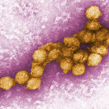
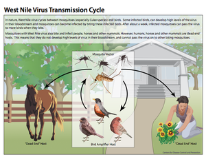
Viruses as a whole are widespread and diverse, especially because they are transmitted via many vectors and cause a variety of symptoms. Many dangerous viruses are transmitted via insects (arthropods) such as mosquitoes. The most common arbovirus, meaning arthropod-borne virus, worldwide is West Nile Virus (WNV), and recent epidemics have brought this virus into the global spotlight as an emerging threat requiring immediate attention for future prevention (Campel et al. 2002).
West Nile virus (Flavividae) is a member of the family Flavivirus and is in the same serocomplex (antigen group) as Japanese encephalitis virus, St. Louis encephalitis virus, and Murray Valley encephalitis virus (Sejvar et al. 2006). Other well-known flaviviruses from this family include Yellow Fever and Dengue Fever, which are also mosquito-borne pathogens (CDC, Flavivirus). West Nile Virus was first identified in the United States in New York in 1999, and since this introduction, it has spread all across the lower 48 states, becoming endemic. Cases of West Nile Virus are particularly concentrated in the Midwestern and Rocky Mountain states during the mid to late summer; however incidences have appeared all across the country year round (Nasci et al. 2103).
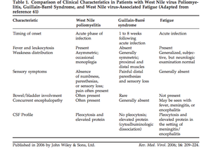
Patients infected with West Nile develop a wide range of symptoms, though in most cases no symptoms arise at all. Out of those infected with West Nile virus, 70-80% are asymptomatic and approximately one quarter develops West Nile fever, which results in a wide spectrum of relatively minor symptoms (Sejvar et al. 2006). However, in rare cases (less than 1%) certain people develop severe neurologic disease, which includes encephalitis, meningitis, and acute flaccid paralysis (Burton et al. 2004). These neurologic disorders tend to be very dangerous and carry a relatively high fatality rate of 10% (Sejvar et al. 2005).
Current research focuses on preventing future epidemics as well as the development of a vaccine for West Nile Virus. In the meantime, efforts to survey outbreaks as well as limit human exposure are utilized to prevent possible outbreaks of the disease. Emerging research also continues to explore the fatal neuroinvasive diseases associated with West Nile Virus in the attempts to prevent and eradicate these as well.
