Bacillariophyta
A Microbial Biorealm page on the phylum Bacillariophyta
Classification
Higher order taxa:
Eukaryota; stramenopiles; Bacillariophtya (including Bacillariophyceae, Coscinodiscophyceae, Fragilariophyceae)
Species:
Thalassiosira rotula, Skeletonema costatum, Thalassiorsira psuedonana
Description and Significance
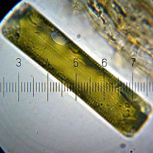
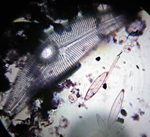
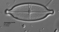
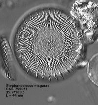
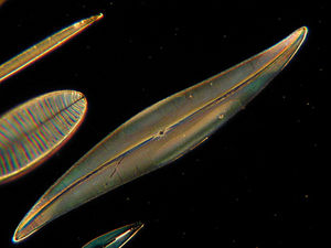
Diatoms are unicells that share the feature of having a cell wall made of silicon dioxide. This opaline or glass frustule is composed of two parts (valves), which fit together with the help of a cingulum or set of girdle bands. These valves have minute holes that allow the exchange gases, nutrients and wastes with the enviroment. Diatoms generally are formed in two general shapes, the centric diatoms, or round, and the pennate diatoms, which are bilaterally symmetrical. These are shown in the pictures at the bottom of the page. On the left is a pennate diatom and on the right a centric.
Diatoms are part of the phytoplankton that are feed on by many marine and freshwater organisms. Their cell wall is not soluble in water so most of them end up on the bottom of lakes and ponds. This sediment will build up and become fossilized rock. This rock is used commercially for everything from abrasives to paper to fertilizer. There are about 10,000 different species of diatoms.
Genome Structure
The U.S. Department of Energy did the first research on a genome for diatoms on the species Thalassiosira pseudonana. It showed, after detailed DNA analysis that diatoms evolved when a heterotroph, a single-celled microbe, engulfed a kind of red alga. The two became one organism, a process called endosymbiosis, and swapped genetic material creating a new hybrid genome. Thalassiosira pseudonana has 34 megabase pairs which encode approximately 11,400 proteins also 129,00 base pair plastid and 44,000 base pair mitochondrial genomes.(For information on the genome project click here.)
Cell Structure and Metabolism
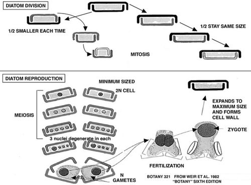
The life cycle of diatoms is shown here:
Diatoms are set up with a cell wall made up of silica and the diatom itself is a single-celled photosynthetic protist. Very little is known about how the cell wall is made, but scientists are researching it to hopefully find a way to reproduce the thin glass-like wall for nanotechnology. Diatoms are autotrophs, which means that they are able to produce their own sugars, lipids and ameno acids. During asexual reproduction, the diatom cell size progressively decreases as each valve produces a smaller complementary valve. When the smaller valves have completely formed the two cells must divide; but because one side of the the diatom is smaller, when they split one is the same size as the original, while one is smaller yet. When they shrink to a certain size, they have to reproduce sexually. To do this they develop an auxospore which then will become a diatom (see diagram above).
Ecology
Diatoms live in a variety of environments, from salt to fresh water (they are even found in moist soil and mosses), a and a wide range of pH levels, temperatures and organic pollution. This variety of living conditions can help tell pollution or other ecological levels of the water. They also vary in their lifestyle, living singly or in a colony.They do not always float freely in the water, they will attach themselves to a rock or another animal in the water. Diatoms may just seem like they are just part of the plankton that feed that fish and animals. But they are huge contributor to the oxygen that is put in to the water and the carbon. It is estimated that 40%, 50 billion to 55 billion tons, of all organic carbon fixation on the planet (transformation of carbon dioxide and water into sugars, using light energy) is carried out by diatoms. This is comparable to all of the world's tropical rainforests.
References
Ben Waggoner. University of California, Berkeley. Diatoms: Life, History and Ecology.
Christopher D. Clack, Eileen Cox, Katie Bantley.. Diatom Morphogenesis. 1994.
Jacob Feldman. Bacillariophytes - Diatoms.
Marine Diatoms. Microscopy UK.
The Micropaleontology Society. Diatoms: Algae with Animal Traits. Science, 1 Oct., 2004.
Owen Davis. University of Arizona. Micropaleontology. April 2003.
Staff, Star Tribune. Species list. June 11, 2004.
