Clostridium botulinum Neurotoxins
Introduction
By Rebecca Dann
Clostridium botulinum is a gram-positive, rod-shaped bacterium and pathogen that is prevalent in marine and soil environments around the world (Figure 1).
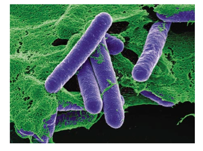
As obligate anaerobes, Clostridium botulinum must live in low oxygen habitats, as higher concentrations are toxic to the cells. These bacteria live in relatively neutral environments and have the most successful growth rates in a pH ranging from 4.6-7.0. Clostridium botulinum is most commonly found as an inactive spore in the shape of an oval. The spores generate a tough outer protective coating and several layers of membranes to enclose the cell and keep it alive. Most Clostridium botulinum spores reside on the surfaces of fruits, dairy products, vegetables, seafood, and various canned foods. As spores, the bacteria usually remain relatively harmless, but when they are activated and resume growth, the bacteria release several different types of potent neurotoxins that can cause the paralyzing disease botulism. The production of the neurotoxins acts as a defense mechanism for the bacteria for protection from intense heat, increased acidity, and possible fragmentation and damages. Clostridium botulinum can produce up to seven different types of toxins named with the letters A-G. The neurotoxins most usually infect individuals by contaminating canned or unrefrigerated food, infecting a wound, or entering a key water source. While all forms of the toxin are destructive, types A, B, E, and F are known to specifically cause botulism in humans, while types C and D are associated with animal botulism. Types A and B mainly cause infant botulism, but types C, E, F, and G have also been identified as causative agents. The factors that regulate and initiate the production of the genes encoding for these neurotoxins are still not known. Researchers are also still in the process of studying how the neurotoxins are released from the bacterial cells.
Cell Structure
Clostridium botulinum is a spore-forming, gram-positive firmicute. These bacteria have the ability to form spores even when in a thriving environment. The formation of spores does not serve the sole purpose of protecting and storing the cell, but it also provides the means for the bacteria to release neurotoxins when they begin germinating. The factors to initiate germination greatly vary and depend on where the spore is located (Webb et al. 2011). In many food products, spores are activated by salt and pH concentrations, or by the addition of chilled or heated temperatures for either storage or preparation. A study by Webb et al. demonstrated that the rate and amount to which spores germinate is directly related to the proximity of spores to each other. The larger the density of spores in the same location, the more germination will occur (Webb et al. 2011). Through designed experiments, a model could be generated to predict the growth rate of the bacterium and determine what conditions initiate the activation of a spore (Webb et al. 2011). This could lead to the discovery of improved prevention techniques to ensure that food is safe and free of Clostridium botulinum.
Genome Structure
This species can be categorized into four genetically and physiologically distinct groups that all produce different types of neurotoxins. The Clostridium botulinum in Group I produce the toxin types A, B, and F. They are proteolytic and have an optimal growth temperature of 37 degrees Celsius but can thrive in temperatures ranging from 12.8-48 degrees Celsius (Peck et al. 2010). This group of toxins most commonly causes infant botulism and contaminates food. Group II consists of Clostridium botulinum that produces type B, E, and F toxins and is also mostly found in contaminated food products. They are nonproteolytic and develop at a lower optimal temperature of 25 degrees Celsius. Group III contains toxins type C and D and are most commonly associated with botulism in animals. Group IV produces toxin type G. The genome for the group I bacteria has been fully sequenced revealing that this bacteria contains chromosomal DNA and a bacteriocin-encoding plasmid (Figure 2).

The chromosomal DNA has 3,886,916 base pairs, which carries 3,650 genes, and the plasmid is composed of 16,344 base pairs, and encodes for 19 genes (Sebaihia et al. 2007). The genes encoding for the potent neurotoxins are located in a cluster on either the main chromosomal DNA or on the large adjacent plasmid (Peck et al. 2010). A significant part of the DNA encodes for the potent neurotoxins, while another large proportion of the genome codes for proteases that are used for the metabolism of proteins. The sequencing of this genome revealed that there has been no recent integration of new foreign DNA. This suggests that this species is incredibly stable and lives in a nonthreatening environment, as no new genes were required for increasing protection, enhancing metabolism, or increasing movement to or detection towards food sources (Sebaihia et al. 2007).
Metabolism
Clostridium botulinum can fully metabolize amino acids and chitin, and can partially metabolize several other polysaccharides. Proteases secreted by the cell can cleave surrounding polypeptides so the bacterium can digest smaller molecules (Sebaihia et al. 2007). There is evidence in the genome that this bacteria uses fermentation pathways that utilize a series of coupled oxidation-reduction reactions to acquire energy, where the oxidation of one amino acid is directly coupled to the reduction of another amino acid. Studies have compiled evidence that the amino acid glycine is first reduced by a glycine reductase complex and then oxidized by a glycine cleavage system (Sebaihia et al. 2007). It has been shown that this bacteria ferments glycine, proline, phenylaline, and leucine. The remaining energy is obtained by sugar metabolism. Clostridium botulinum uses the second most abundant sugar, Chitin, as its second main source of energy (Sebaihia et al. 2007). Chitin is commonly found in the exoskeletons of arthropods and insects, such as lobsters and mollusks, and in the cell walls of fungi, both of which are prevalent in the marine and soil environments in which this bacteria resides (Figure 3).

Clostridium botulinum’s genome encodes five different enzymes that are capable of metabolizing this sugar. In addition to being a source of energy, chitin can also provide supplementary carbon and nitrogen (Peck et al. 2010). Starch is a potential source of energy, but these bacteria do not have all of the enzymes required to fully degrade this polysaccharide. Several types of Clostridium botulinum have a complete glycolysis system in addition to having the capability of fermentation (Sebaihia et al. 2007).
Formation of Endospores
A key characteristic of Clostridium botulinum is their ability to form dormant spores that can withstand extreme conditions, such as high pressure, UV light, and heat treatments. Sporulation is the reason for why this bacterium is such a food hazard and poses such a threat to those who are exposed to the species. To ensure that food is free of proteolytic botulinum, it must be treated with a temperature of at least 121 degrees Celsius for a minimum of three minutes; this process has been termed the “botulinum cook.” Spore germination in proteolytic Clostridium botulinum is initiated by the presence of the amino acid L-alanine, which activates germinant receptor proteins located in the inner membrane of the spore. These proteins are encoded by three germinant receptor operons that are expressed during germination (Peck et al. 2010).
Spores of the non-proteolytic Clostridium botulinum are less heat resistant in comparison with the proteolytic bacteria. A temperature range of only 80-85 degrees Celsius for several minutes prevents spore germination. Unlike the proteolytic bacteria, this strain of botulinum can be activated by the presence of a lysozyme, an enzyme that is prevalent in common foods. The lysozyme can enter the protective spore coat and hydrolyze the peptidoglycan layer of the cortex (Peck et al. 2010). This allows the core, which contains the bacteria’s DNA, to be exposed to its surroundings, which activates germination. Spore germination in non-proteolytic botulinum can also be initiated by the presence of two amino acids, such as L-alanine and L-lactate. The germinant receptor proteins that allow the amino acids to initiate a response are only encoded by one germinant receptor operon instead of three as in the proteolytic botulinum (Peck et al. 2010).
Pathology
The discovery and detection of new emerging neurotoxins found in contaminated food has fueled a growing concern for ensuring that safe food is distributed in numerous countries around the world. Many recent studies have investigated new techniques in which to regulate Clostridium botulinum growth and diversification. This requires improved mechanisms of detection of this bacteria and a comprehensive understanding of how and why it flourishes in the environments that it does and how exactly it causes such a biological response. Even though reported cases of botulism around the world are relatively infrequent, these bacteria are still a hazard. The smallest microscopic amounts of the neurotoxins released by these bacteria can cause the most damaging form of the disease. It has been reported that only 30 ng of the neurotoxin can induce a drastic effect (Sebaihia et al. 2007).
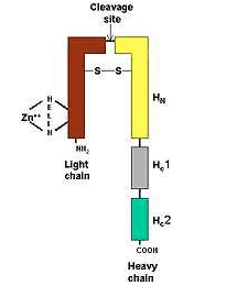
There is currently little known about the genes that encode for the neurotoxins and the assembly and production of the molecule. Numerous studies have yielded inconsistent results, but the structure of the neurotoxin has in fact been discovered. The toxin is in the form of a noncovalently bound complex that contains several nontoxic proteins that consist of hemegglutinin and nonhemegglutinin, and weighs 150 kDa (Sebaihia et al. 2007). In order for this polypeptide molecule to become toxic, it is cleaved by a protease at one-third the distance from the N terminus. The exact enzyme that performs this function has still yet to be been determined. This action yields two fragments: a smaller, lighter fragment weighing 50 kDa and a heavier fragment with a larger weight of 100 kDa (Figure 4). These two fragments are kept joined together by a disulfide bond and collaborate to produce the damaging biological response (Todar 2009).
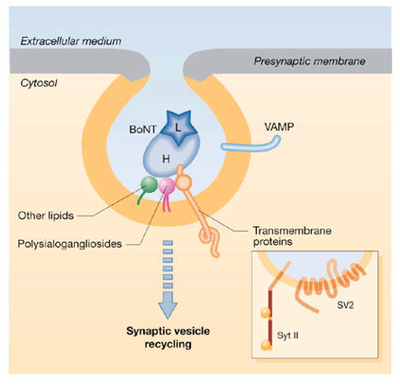
The neurotoxins target the body’s peripheral nervous system, which is greatly exposed to pathogens, as it is not protected by the blood-brain barrier or bone. They pass through the membrane and enter into the neuron cell by endocytosis. Pirazzini et al. hypothesized that the heavier chain forms a channel within the membrane in which the lighter chain can then pass through into the cytoplasm (Pirazzini et al. 2013). They found evidence that the heavier chain contains two polysialoganglioside binding sites that allow the toxin to successfully bind to the membrane and use the low pH environment on the outside of the cell to drive its entry into the neuron which has a neutral concentration (Figure 5). study also discovered that 37 degrees Celsius is the optimum temperature for the transport of the toxin through the plasma membrane, as the translocation of the lighter chain into the cell occurred in just minutes, which is an extremely rapid pace (Figure 6).
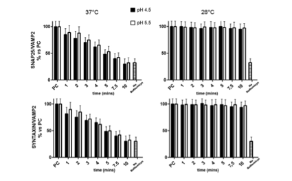
When the Pirazzini et al. tested the effects of 20 degrees Celsius on toxin transport they found that no toxins entered the neuron. These results indicate that transfer of the toxins through the membrane is greatly dependent on temperature.
Once inside the neuron, the toxin binds to the presynaptic membrane of the cholinergic nerve terminals, blocking the release of the neurotransmitter acetylcholine. Acetylcholine plays an essential role in the body, as it is responsible for regulating the somatic nervous system, which controls the voluntary movements of the skeletal muscles, and is the only of its kind. No other neurotransmitter initiates this type of movement. As a result, if the toxin does in fact bind to the receptor it has very damaging effect, as this blocking mechanism prevents the nervous system from communicating with the muscles, resulting in limited muscle movement and paralysis.
The botulinum toxin produces specified cleaving proteases that allow the pathogen to successfully attach to the synaptic vesicles. Studies have identified the synapse as the synaptic vesicle protein SV2 (Peng et al. 2010). If this specific receptor SV2 is not present within a cell then the neurotoxin does not produce the same effect. Polysialogangliosides and SV proteins surround the membrane to facilitate the binding of the neurotoxin (Figure 7).
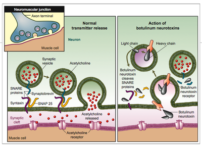
The carboxy-terminal domain of the heavy chain recognizes a specific binding site, while the nitrogen-terminus transports the lighter chain into the nerve cytosol (Peck et al. 2010). The lighter chain contains metalloproteases that target specific proteins involved in controlling the exocytosis machinery (Verderio et al. 2006). The inhibition of this integral machinery stops the release of acetylcholine and the neuron fails to send an important signal throughout the body. The lighter chain also decreases the stability of the binding complex, further preventing acetylcholine from being able to bind to the synaptic vesicles (Peck et al. 2010).
In addition to releasing neurotoxins when exposed to varying environmental conditions, Clostridium botulinum also increases production of proteases that are secreted from the cell to breakdown polypeptides to contribute to contaminating food and therefore increasing its own toxicity. A large proportion of the bacteria’s genome encodes for several different variations of protease enzymes (Sebaihia et al. 2007).
Infant Botulism
Infant botulism occurs when the botulinum heat-resistant spores are consumed and germinate within the human intestine (Peck at al. 2010). It appears in infants less than one year old, and is caused due to the lack of microflora in an infant’s intestines. The lack of developed microbial communities can leave a newborn unprotected when encountering foreign bacteria ingested from unknown foods. Without the complete microflora, the Clostridium botulinum spores are able to germinate, which releases the threatening neurotoxins. Type A and B toxins are most commonly released, but types C, G, F and E have also been witnessed to cause infant botulism (Nevas et al. 2005).
The most commonly contaminated food that carries infant botulism is honey. The number of spores inhabiting a sample of honey was found to range from 1-60, but even a small number of less than ten spores are still more than enough to produce an overwhelming amount of neurotoxins (Nevas et al. 2005). The spores are only activated when they come in contact with the environment of the intestines, as the conditions created by honey keep the spores in an inactive state. Honey maintains a relatively anaerobic environment due to the viscosity of the substance and retains a low pH. It also has an extremely high sugar concentration and a low protein concentration, which could be harmful, as Clostridium botulinum relies on amino acids as an essential nutrient for energy and for initiating germination. Maintaining spore form can be viewed as beneficial, as it does not allow for the release of harmful toxins in a prevalent food, but it could also be seen as potentially damaging, since the spores can remain in the honey samples for theoretically as long as they can maintain their spore form (Nevas et al. 2005). As a result, the Clostridium botulinum could survive in honey samples for years undetected creating a potential threat for an outbreak of infant botulism.
The results of an experiment conducted by Nevas et al. testing the concentration and type of Clostridium botulinum in honey samples from different northern European countries concluded that toxin types B and A were most prevalent in the Danish and Norwegian honey samples, while only type E neurotoxin was found in the Swedish honey samples (Nevas et al. 2005). Overall, there was more bacterium present in the Danish honey than in the other samples measured from surrounding countries. Nevas et al. hypothesized that the Clostridium botulinum spores could have originated from a nearby water source that came in contact with the honey samples. This would suggest that concentration of bacterial spores in honey reflects the total concentration of bacteria present in the surrounding natural environment. This would imply that the soil and aquatic environments in Denmark contain the most of these harmful bacteria.
Food-borne Botulism
Food-borne botulism is caused by the consumption of food products that already contain the botulinum toxin in its potent non-spore form. Clostridium botulinum toxins have mostly been found to contaminate raw meat and canned and bottled goods that have extended their shelf-life (Peck et al. 2010). The most common method in which to rid of these toxins is to always heat chilled canned food before it is consumed. Outbreaks of food-borne botulism have been reported all over the world, with the first official case investigated in 1895 involving contaminated blood sausages. Both proteolytic and non-proteolytic Clostridium botulinum can cause food-borne botulism. The proteolytic bacteria most commonly release either the type A or type B toxin and are found in canned foods, while the non-proteolytic Clostridium botulinum produce the type A or type E neurotoxin and are present in meat, fish, or homemade meals (Peck et al. 2010).
Smelt et al. conducted an experiment that attempted to discover or gain better insight into how non-proteolytic Clostridium botulinum can grow and develop at refrigerated temperatures (Smelt et al. 2013). This experiment would be especially beneficial for keeping dairy products safe from the bacteria’s neurotoxin. It had been previously determined that heated spores display a much different development process than unheated spores. The results of this experiment showed that there were less spores present in the colder samples, but the cold temperatures did not cause this result, as the cold-grown bacteria had growth and replication rates in normal ranges (Figure 8).
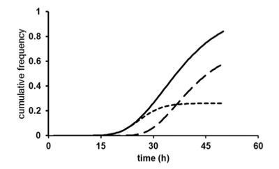
The colder temperature could have influenced other conditions, such as oxygen levels or the type of media used for the culture (Smelt et al. 2013). 90% of the spores treated with a heated condition could not germinate, and a considerable proportion could not replicate. While this study was not able to generate any conclusions as to why cold-grown botulinum was found in smaller samples but retained a normal growth rate, it did demonstrate that Clostridium botulinum could survive in colder conditions. This implies that even refrigerated food can contain this bacterium.
Forms of Prevention and Treatment
High-pressure thermal treatments are most commonly used as a remedy to remove all of the harmful bacteria, even when they are still in the form of a spore. If the spores are treated with heat of 100 degrees Celsius for over an hour, the spores are inactivated and unable to produce toxins (Pirazzini et al. 2013). Cooking has become an easy prevention technique to ensure safe preparation of food. Maintaining a low pH below 4.6 also prevents the growth of these bacteria. While food corporations have used these methods for many years, new cases where certain food products or sources are contaminated continue to arise, and novel mechanisms for regulation must be tested.
If an adult individual is already intoxicated by the botulinum neurotoxin, equine antitoxin can be administered to help rid of the symptoms. The drug inhibits the neurotoxins that have yet to bind to the nerve receptors from doing so, decreasing the toxin’s ability to cease muscle movement. Some cases of botulism can take months to years for a full recovery, and fatality occurs in approximately 5-10% of the cases reported (Peck et al. 2010). The cost to treat this devastating disease can be very expensive as it can be a long process. Cases of infant botulism can be treated with Botulism Immune Globulin Intravenous, which provides essential fluids and nutrients.
Conclusion
While there are currently successful methods to rid of Clostridium botulinum from contaminated food sources, further research is still needed to ensure that this bacteria presents no threat, as botulism is one of the most devastating diseases. By gaining a more comprehensive understanding of the structure of the Clostridium botulinum neurotoxins and the mechanisms for why and how they are synthesized by the bacteria can help generate ideas as to how to cure this disease and prevent it from even occurring.
