Xenorhabdus nematophilus
Classification
Bacteria; Proteobacteria; Gammaproteobacteria; Enterobacteriales; Enterobacteriaceae;Xenorhabdus
Species
Xenorhabdus nematophilus Taxonomy
Introduction
Xenorhabdus nematophilus is a gram-negative bacterium in the family Enterobacteriaceae. This microbe can be described as entomopathogenic (i.e. an organism which can kill arthropods by poisoning with its own toxins or those it harbors).
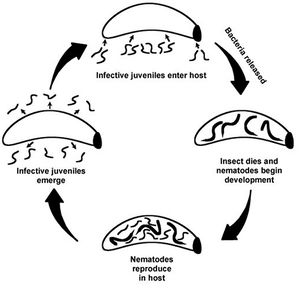
X. nematophilus is not found free living in the soil environment. It exists in a symbiotic relationship with insect-parasitizing nematodes of Steinernema carpocapsae . The interaction is specific to each species. They are found ubiquitously in soil environments. Their ecological significance is particularly apparent in agriculture, as a form of biological control of pest insect species. The biological processes of the bacterium are matched by the needs of the nematode and vice versa. Together this mutualistic relationship results in the predation of insect species, such as those in the order Lepidoptera.
Important Morphology
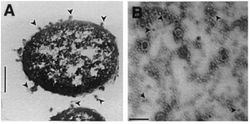
Gram-negative bacteria produce two-layer outer membrane blebs. As outer membrane vesicles, these spherical structures encapsulate toxic proteins to protect them from the degradation by host enzymes. X. nematophilus excrete blebs into its growth medium [SOURCE:khandelwal]. The vesicles have electron-dense contents and also contained proteins. The excretions have properties of insecticides, antimicrobial agents, and degradative enzyms. [KHAND, SOURCES]
Life Cycle
The life cycle of this microbe has been described as having two phases, where different metabolites are secreted. Xenorhabdus nematophilus is considered stationary once it has been regurgitated by the nematode into the hemocoel of the larval host and infects and kills it. At the different phases of X. nematophilus, the excretions differ in their virulent properties. During the stationary phase, the cells produce proteases, phospholipases, antibiotics, and inclusions composed of crystal proteins. These all function to facilitate an optimal growing environment for both the itself and the nematode. Research has found that certain phenotypic traits are common to all X. nematophilus cells, while virulence and motility are variable and can be influenced by environmental conditions. Motile X. nematophilus has petritrichous flagella.
Biological interaction

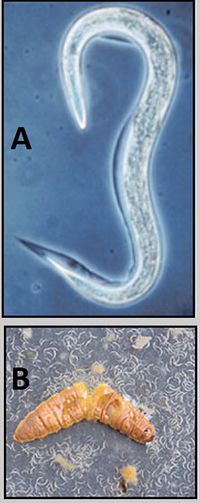
The interaction between Xenorhabdus nematophilus and Steinernema carpocapsae is a specific mutualism. The bacterium is contained in a vesicle of the foregut of an infective juvenile stage soil nematode. Once the nematode finds and enters an insect host, the bacterium in released into the hemocoel of the insect.
Why S. carpocapsae needs X. nematophilus
The nematode is not a free living organism and requires a host for its life cycle to be completed. Only the infective juvenile stage of S. carpocapsae is able to move through the environment in order to find a new host. In order to create an optimal growing environment in which to grow and reproduce, the nematode needs the entomopathogenic bacterial symbiont. X. nematophilus will secrete compounds that kill the insect host (found to be toxic proteins). Additional compounds include enzymes that bioconvert the insect host. Antimicrobial agents are also secreted that protect the insect carcass from putrification from other microbes. [Khandelwal, Floyd]. In the initial stages of infection of the insect host, X. nematophilus inhibits the growth of various fungal and bacterial competitors. The metabolites exuded by the bacterium are known to have antifungal, nematicidal, or insecticidal effects. X. nematophilus limits competition so that the nematode and itself can use the insect larvae carcass nutrients.
Why X. nematophilus needs S. carpocapsae
The bacterium X. nematophilus is not found in the soil environment. It cannot survive in water or soil for long by itself, as it is an endosymbiont of nematodes (Photorhabdus luminescens has the same characteristic).
S. carpocapsae facilitates the death of the larval host. The nematode is only free living during its infective juvenile stage, and it is at this time it can disperse through the soil environment to locate a larval host. This function has allowed both organisms to become ubiquitous in nearly all soil environments.
When a host is found, S. carpocapsae enters the body cavity of the larvae. Once it is in the hemocoel of the insect, the X. nematophila are released from the gut of the nematode.The hemocoel is a series of interconnected tissue spaces through which blood circulates in arthropods, thus it is an ideal structure to start an infection.
How interaction influences community
Populations of S. carpocapsae and X. nematophilus influence the populations of their insect hosts. This symbiotic relationship causes mortality of the host in it’s larval stage. Most commonly Lepidopteran species are the target host, and their larvae function most importantly as consumers of plants. In the case of agriculture, there are many lepidopteran species that are pests to the crop. For example, the Helicoverpa armigera (corn ear worm). In a study of the toxicity of X. nematophilus on H. armigera larvae, it was found that LD50= 4x10⁴ cells/gram of diet is the dosage that will cause the death of 50% of the test population. Non-fatal levels of infection resulted in symptoms such as small size and moribund (i.e. near death). These results are true for when the bacterium invades the hemocoel of the larva or is ingested orally. [SOURCE KHANDEL]
Interactions with other species 5
potato
Phtytophthora infestants oomycete negative/positive?. outcome? “”However, earlier identified soluble antibiotics, such as xenorhabdins, indole compounds (McInerey et al., 1991a, b) and nematophin (Li et al., 1997) from other Xenorhabdus spp. have shown antifungal activity against soil fungi.""
fungi
X. nematophilus has antagonistic interactions with entomopathogenic fungi.
Niche
Xenorhabdus nematophilus is ubiquitous across all habitat types. Since it has been shown that Steinernematid (and Heterorhabditid) nematodes are exclusive to soil environments, and they have been isolated from every continent, excluding Antarctica. Entomopathogenic nematodes exist in a diverse range of soil habitats including farmland, forests, beaches, and deserts. A survey of entomopathogenic nematodes had confirmation of the nematodes in 2-25% of the sites sampled.
Cultivated Fields1
Subsection 2
Subsection 2
Key Microorganisms, Microbial Communities

The outer membrane of gram-negative bacteria performs many functions. The membrane may be specialized to increase survival in adverse conditions. The outer membrane has a mechanism for protein secretion, that in the case of bacterial pathogens will kill the host. The bacteria can then utilized the the nutrient rich source of the host cadaver. Virulence factors are contained in outer membrane vesicles and secreted to the environment. Pseudomonas aeruginosa, Proteus mirabilis, and Serratia marcescens are other examples of bacteria that package enzymes such as proelastases, hemolysins, and protease in vesicles. The Bacteroides gingivalis has similar vesicle properties to X. nematophilus where it releases toxins from its outer membrane.
Other Microbes
Photorhabdus luminescens is related to X. nematophilus. They were found to have homology of genes that code for the insecticidal proteins associated with nematodes. P. luminescens also has oral insecticidal activity.
Insect function
The most common hosts for the symbiotic pair are species in the order Lepidoptera. Lepidoptera have numerous roles in an ecosystem. Examples of species mentioned in the sources include: Helicoverpa armigera (corn earworm) is a moth whose larvae feed on a wide range of crops; Pieris brassicae (cabbage white butterfly) larvae are also crop pests; Manduca sexta (greater wax moth); and Galleria mellonella is a moth that is a pest of honey bees combs and fruit.
Functions of the larval forms of these species include damage to plant species from feeding on the tissues. Damage to crops can reduce yield. Insects can also act as vectors for other diseases. Adult forms of these species can serve as pollinators or a food source for other organisms in the community.
other
potato/fungi Phtytophthora infestants
Microbial processes
Bacterium Functions
Toxic proteins
morgan et al. there is a DNA region of Xn found to encode for insecticidal protein, chitinase, and other pathogenic attributes. This suggests that the toxins of Xn are made up of multiple components. (multiple polypeptides)
TOXINS: the particulate fraction of the insecticidal proteins are more active than the soluable fraction (high-speed centrifugation). "Heating OMVs at 80 C for 15 min inactivated the insecticidal activity"When treated with a proteinase, up to 80% of the OMV proteins were degraded and inactive.
Outer Membrane Vesicles
OMV emanate from the cell, encapsulate toxic proteins- protects proteins from degradation by the cells enzymes. vesicles are like a delivery system for the cell. separated from the cytosol and in most organisms is separated by at least one lipid bilayer. During growth, OMV are sloughed off from the surface of a cell. OMVs for Sn contain electron-dense compounds, proteins, porins, Abiosis excretions the bacteria produce antibiosis in vivo (in an organism) and in vitro (in artificial) Nutrient excretions
Nematode Functions
Nematodes are classified as part of the microfauna in a soil environment.Food web studies show that fauna contribute up to 30% of the total net nitrogen mineralization, mainly from microbial-feeding nematodes and protozoa. At this level of the soil ecosystem, nitrogen and other nutrients that would otherwise be locked up in organic form, are released through the decomposition activities of the microfauna. The possible Net Primary Productivity of the system is increased. Specific to the interaction between X. nematophilus and S. carpocapsae, the insect carcass is consumed by the nematode and the bacterium, efficiently using all available nutrients. The symbiotic relationship between the two organisms allows for maximum uptake or release of nutrients. Termite have microbial symbionts in the termite gut, and together the two organisms decompose ligno-cellulose.
Current Research
Biological Control
Agriculture. the most successful insecticide based on a microbe is that from the bacterium Bacillus thuringiensis. issue: resistence. so look for new genes. The genes must be orally active protein toxins. Such that the gene can be genetically engineered to be expressed in the plant tissue eaten by the pests. This is important because then only the insect species that are damaging the crops will be killed. This allows for safer pesticide use in cultivated fields, and it allows for greater biodiversity.
In Agricultural systems. The use of X. nematophilus as a bio-pesticide is limited. It cannot be stored indefinitely on a shelf under any conditions. For the bacterium to be effective, it needs to be transported and used within a time frame. And as shown by research done on outer membrane vesicles, it cannot be exposed to excessive heat. This can be a topic of further research. LINK.
What factors allow for the bacteria to recognize environment?
The bacteria must have some mechanism by which to recognize whether it is in it’s host environment and regulate gene expression accordingly. ===What are the specific functions of the proteins in the outer membrane vesicles? unable to determine the toxicity individual proteins present in OMV mixtures for Xn. They confirmed that the OMV contain insecticidal protein toxins. Chitinase is likely to be an OM protein, but its effectiveness is not known. The presence of chitinase increases the pathogenic possibilities of the OMV compounds for Xn.
References
1. Floyd L. Inman and Leonard Holmes, 2012. Effect of Heat Sterilization on the Bioactivity of Antibacterial Metabolites Secreted byXenorhabdus nematophila. Pakistan Journal of Biological Sciences, 15: 997-1000.
1. H. Fossing, V.A. Gallardo, et. al. "Concentration and transport of nitrate by the mat-forming sulphur bacterium Thioploca". Nature. 1995. Volume 374. p.713-715. 2. Codispoti, L.A. et al. "High Nitrite Levels off Northern Peru: A Signal of Instability in the Marine Denitrification Rate". Science. 1986. Volume 233. p.1200-1202. 3. Schulz, H., Jorgensen, B., Fossing, H., and Ramsing, N. "Community Structure of Filamentous, Sheath-Building Sulfur Bacteria, Thioploca spp., off the Coast of Chile". Applied and Environmental Microbiology. 1996. Volume 62. p. 1855-1862. 10. Teske, A., Ramsing, N. B., Kuver J., and Fossing, H. "Phylogeny of Thioploca and related filamentous sulfide-oxidizing bacteria". Syst. Appl. Microbiology. 1996. Volume 18. p. 517-526. 11. Larking, J., and Strohl, W. "Beggiatoa, Thiothrix, and Thioploca". Ann. Rev. Microbiology. 1983. Volume 37. p. 361-362. 12. Schulz, Heide, and Schulz, Horst "Large Sulfur Bacteria and the Formation of Phosphorite". Science. 2005. Volume 307. p. 416-418.
Edited by <Chloe M. Mattia>, a student of Angela Kent at the University of Illinois at Urbana-Champaign.

