Pneumococcal meningitis and the role of Streptococcus pneumoniae: Difference between revisions
Andersonam (talk | contribs) No edit summary |
Andersonam (talk | contribs) No edit summary |
||
| Line 16: | Line 16: | ||
<br><b>Superscript:</b> Fe<sup>3+</sup> | <br><b>Superscript:</b> Fe<sup>3+</sup> | ||
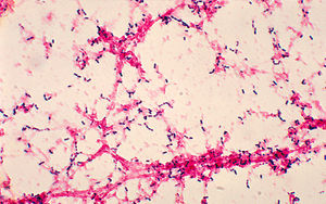
[[Image:phil-2896.jpg|thumb|300px|left|Figure 2. Photomicrograph of gram-positive <i>Streptococcus pneumoniae</i> grown from blood culture, on 1978. By Dr. Mike Miller, at the [http://www.cdc.gov/ CDC].]] | [[Image:phil-2896.jpg|thumb|300px|left|Figure 2. Photomicrograph of gram-positive <i>Streptococcus pneumoniae</i> grown from blood culture, on 1978. By Dr. Mike Miller, at the [http://www.cdc.gov/ CDC].]] | ||
<br> | |||
<br> | |||
[[Image:surface-proteins.jpg|thumb|500px| | <br> | ||
<br> | |||
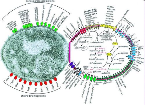
[[Image:surface-proteins.jpg|thumb|500px|right|Figure 3. Depiction of <i>Streptococcus pneumoniae</i> substrate transport, carbohydrate and glutamine metabolism, and selected categories of cell surface proteins, for full caption see paper Hoskins, et. al 2001 [http://www.ncbi.nlm.nih.gov/pmc/articles/PMC95463/figure/F1/ PubMed].]] | |||
<br>Introduce the topic of your paper. What microorganisms are of interest? Habitat? Applications for medicine and/or environment?<br> | <br>Introduce the topic of your paper. What microorganisms are of interest? Habitat? Applications for medicine and/or environment?<br> | ||
Revision as of 00:31, 20 April 2015
Introduction

By Avery Anderson
At right is a sample image insertion. It works for any image uploaded anywhere to MicrobeWiki. The insertion code consists of:
Double brackets: [[
Filename: PHIL_1181_lores.jpg
Thumbnail status: |thumb|
Pixel size: |300px|
Placement on page: |right|
Legend/credit: Electron micrograph of the Ebola Zaire virus. This was the first photo ever taken of the virus, on 10/13/1976. By Dr. F.A. Murphy, now at U.C. Davis, then at the CDC.
Closed double brackets: ]]
Other examples:
Bold
Italic
Subscript: H2O
Superscript: Fe3+


Introduce the topic of your paper. What microorganisms are of interest? Habitat? Applications for medicine and/or environment?
Pathogenesis
Include some current research, with at least one figure showing data.
Risk Factors
Include some current research, with at least one figure showing data.

Treatment
Include some current research, with at least one figure showing data.
References
[1] Hodgkin, J. and Partridge, F.A. "Caenorhabditis elegans meets microsporidia: the nematode killers from Paris." 2008. PLoS Biology 6:2634-2637.
Authored for BIOL 238 Microbiology, taught by Joan Slonczewski, 2015, Kenyon College.
