Zaire ebolavirus: Pathogenesis, History, and Treatment of the Deadly Virus: Difference between revisions
| Line 36: | Line 36: | ||
[[Image:MB-003.png|thumb|300px|right|The results of MB-003 testing. MB-003 recipients were compared against control and history control groups. The survival rate is compared against the days after exposure to the virus.http://stm.sciencemag.org/content/5/199/199ra113.full]] | [[Image:MB-003.png|thumb|300px|right|The results of MB-003 testing. MB-003 recipients were compared against control and history control groups. The survival rate is compared against the days after exposure to the virus.http://stm.sciencemag.org/content/5/199/199ra113.full]] | ||
== | ==Virus Structure== | ||
The genome of the Ebola virus isn’t very complex, only containing enough genetic information to code seven to eight proteins. The virus is made of RNA, glycoproteins, a membrane, and matrix proteins designed to help the virus keep its shape (Goodsell). | |||
The way the virus gets its genome into the host cell is via glycoprotein. The glycoprotein binds to receptors of the host cells. The protein extends from the virus and is covered in carbohydrate chains, which help the virus hide itself from the host’s immune system. The glycoprotein also functions as a viral fusion protein. The glycoprotein binds to the host cell where it changes shape. This change quickly pulls the virus and the host cell very close together, rapidly, fusing their membranes. This process is very similar to the glycoloprotein of an influenza strain and the glycoprotein of HIV (Goodsell). | |||
Ebola virus is a part of the Filoviridae family of viruses. The size of the virus varies, depending on the strain. The Filoviridae family varies in size with a genome of negative-sense RNA. The length of the genome is approximately 19 kb long. In the RNA, there are seven open reading frames resulting in eight different proteins that have various jobs from matrix proteins to non-structural proteins. These eight proteins are responsible for the function of one of the deadliest viruses known to humans (Falasca et al.). | |||
The genome of the virus is protected by the nucleocapsid. The purpose of the nucleocapsid is to protect the genome until the virus is attached to a host cell, so the genome can be injected into the host, where it can undergo replication and infect the cell. The nucleocapsid of the Ebola virus is made up of nucleoproteins that surround the RNA in a helical fashion. The capsid proteins aren’t incredibly rigid, allowing the virus to have some flexibility. One of the proteins that makes up the capsid is very important not just for structure but also for replication of the genome. This protein is Polymerase (L) Protein. Once the genome is inserted into the host cell, this protein is responsible for the replication of the genome (Goodsell). | |||
The matrix protein in Ebola virus has been named the VP40 protein. The matrix of the virus is responsible for the shape and drives the budding process of the host cell post infection. The protein is also responsible for being a connector between the nucleocapsid and the membrane of the virus, again helping the virus maintain a shape. Matrix proteins commonly have several jobs in the viral process. Ebola matrix protein has three distinct confirmations with three different functions in the cell. One confirmation is a hexamer responsible for priming the RNA to initiate replication once the virus is inside the host cell. The next confirmation is an octamer that is responsible for regulating the transcription of the genome. A final confirmation is a dimer that’s major function is protein transport. (Goodsell) | |||
==Section 4== | ==Section 4== | ||
Revision as of 01:48, 29 April 2016
Section

By Alexander Manning
At right is a sample image insertion. It works for any image uploaded anywhere to MicrobeWiki.
The insertion code consists of:
Double brackets: [[
Filename: PHIL_1181_lores.jpg
Thumbnail status: |thumb|
Pixel size: |300px|
Placement on page: |right|
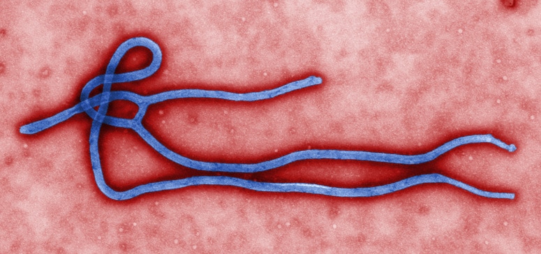
Legend/credit: Electron micrograph of the Ebola Zaire virus. This was the first photo ever taken of the virus, on 10/13/1976. By Dr. F.A. Murphy, now at U.C. Davis, then at the CDC.
Closed double brackets: ]]
Other examples:
Bold
Italic
Subscript: H2O
Superscript: Fe3+
Introduce the topic of your paper. What is your research question? What experiments have addressed your question? Applications for medicine and/or environment?
History of the Virus

The Ebola virus was first discovered in 1976 near the Ebola River in the Democratic Republic of the Congo (Outbreaks Chronology: Ebola Virus Disease). In that year, there were 318 confirmed cases of the virus infecting humans, resulting in 280 deaths. Over the next several years, there were only a few reported cases, usually in controlled situations that didn't result in any fatalities. Then, in 1995, there was an outbreak of the virus in the Democratic Republic of the Congo. This outbreak resulted in 315 confirmed cases with 250 deaths reported. Just five years later, there was an outbreak in Uganda that resulted in 425 cases and 224 deaths. This outbreak was believed to have been a result of the attendance of a funeral. The person being buried was killed as a result of the Ebola virus infection, and the people attending the funeral were not wearing the proper attire to prevent transmission of the virus. The following year, there was another outbreak, this time in the Republic of Congo. This outbreak saw 57 cases with 43 deaths. Six years later in 2007, there were two separate outbreaks. One outbreak was in western Uganda, the other in the Democratic Republic of the Congo. The Uganda outbreak had 149 cases with 37 deaths. The outbreak in the Democratic Republic of the Congo had 264 cases with 187 reported deaths. Seven years later, in 2014, the largest outbreak in history broke out in western Africa. During this devastating outbreak, there were over 27,000 reported cases with over 11,000 reported deaths (Outbreaks Chronology: Ebola Virus Disease). This virus has now been wreaking havoc on the world and western Africa in particular, for four decades now. It has claimed thousands and thousands of victims and has terrified countries into preventing citizens from returning home on the chance that they could spread the virus causing an epidemic that would render their country helpless. Originally discovered a few dozen miles away from a river in the Democratic Republic of the Congo, this virus has since developed a stigma making it known for the brutal death it is capable of causing. Vast amounts of research have been done on the virus. All aspects of the virus have been considered, from function and pathogenesis to treatment and vaccinations.
The Ebola virus is seen as one of the most deadly viruses known to humans with a 50-90% mortality rate. There are currently five known strains of the virus all depending on the location of the virus. However, only four of the five viruses are capable of causing infection in a human host. This fifth virus is capable of infecting primates (CDC).
Virus Entry into the Host
The Ebola virus enters the host through basic entry paths. It can enter through mucous membranes, broken skin, or by parental transmission. One way that the virus is so deadly is its ability to attack almost every type of human cell. It is capable of binding to these membranes of essentially every human cell except for lymphocytes. The entry of the cell is controlled by glycoprotein that is responsible for binding the virus to the cell receptors. Once the virus is bound to the cell receptors, the membranes fuse together, followed by the opening of the capsid injecting the genome of the Ebola virus into the cell. The genome then undergoes transcription and replication. (Goodsell)

Virus Structure
The genome of the Ebola virus isn’t very complex, only containing enough genetic information to code seven to eight proteins. The virus is made of RNA, glycoproteins, a membrane, and matrix proteins designed to help the virus keep its shape (Goodsell).
The way the virus gets its genome into the host cell is via glycoprotein. The glycoprotein binds to receptors of the host cells. The protein extends from the virus and is covered in carbohydrate chains, which help the virus hide itself from the host’s immune system. The glycoprotein also functions as a viral fusion protein. The glycoprotein binds to the host cell where it changes shape. This change quickly pulls the virus and the host cell very close together, rapidly, fusing their membranes. This process is very similar to the glycoloprotein of an influenza strain and the glycoprotein of HIV (Goodsell).
Ebola virus is a part of the Filoviridae family of viruses. The size of the virus varies, depending on the strain. The Filoviridae family varies in size with a genome of negative-sense RNA. The length of the genome is approximately 19 kb long. In the RNA, there are seven open reading frames resulting in eight different proteins that have various jobs from matrix proteins to non-structural proteins. These eight proteins are responsible for the function of one of the deadliest viruses known to humans (Falasca et al.).
The genome of the virus is protected by the nucleocapsid. The purpose of the nucleocapsid is to protect the genome until the virus is attached to a host cell, so the genome can be injected into the host, where it can undergo replication and infect the cell. The nucleocapsid of the Ebola virus is made up of nucleoproteins that surround the RNA in a helical fashion. The capsid proteins aren’t incredibly rigid, allowing the virus to have some flexibility. One of the proteins that makes up the capsid is very important not just for structure but also for replication of the genome. This protein is Polymerase (L) Protein. Once the genome is inserted into the host cell, this protein is responsible for the replication of the genome (Goodsell).
The matrix protein in Ebola virus has been named the VP40 protein. The matrix of the virus is responsible for the shape and drives the budding process of the host cell post infection. The protein is also responsible for being a connector between the nucleocapsid and the membrane of the virus, again helping the virus maintain a shape. Matrix proteins commonly have several jobs in the viral process. Ebola matrix protein has three distinct confirmations with three different functions in the cell. One confirmation is a hexamer responsible for priming the RNA to initiate replication once the virus is inside the host cell. The next confirmation is an octamer that is responsible for regulating the transcription of the genome. A final confirmation is a dimer that’s major function is protein transport. (Goodsell)
Section 4
Conclusion
References
Authored for BIOL 238 Microbiology, taught by Joan Slonczewski, 2016, Kenyon College.
